4L8W

Crystal structure of gamma glutamyl hydrolase (H218N) from zebrafish complex with MTX polyglutamate


Experimental Data Snapshot

  • Method: X-RAY DIFFRACTION
  • Resolution: 2.39 Å
  • R-Value Free: 0.258 
  • R-Value Work: 0.193 
  • R-Value Observed: 0.196 

wwPDB Validation   3D Report Full Report


Ligand Structure Quality Assessment 


This is version 1.1 of the entry. See complete history


Literature

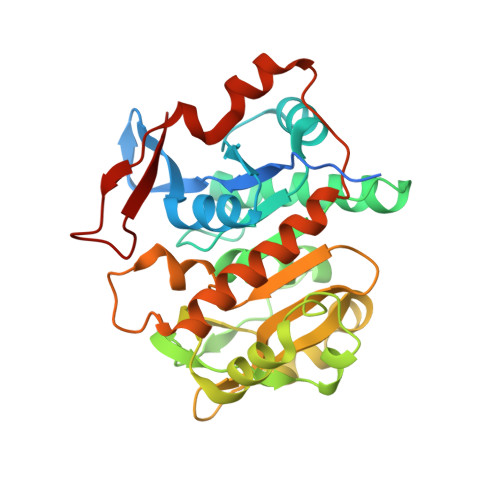
Structural insights into the hydrolysis and polymorphism of methotrexate polyglutamate by zebrafish gamma-glutamyl hydrolase

Chuankhayan, P.Kao, T.-T.Lin, C.-C.Guan, H.-H.Nakagawa, A.Fu, T.-F.Chen, C.-J.

(2013) J Med Chem 56: 7625-7635

  • DOI: https://doi.org/10.1021/jm401013e
  • Primary Citation of Related Structures:  
    4L7Q, 4L8F, 4L8W, 4L8Y, 4L95

  • PubMed Abstract: 

    γ-Glutamyl hydrolases (γGH) catalyze the hydrolysis of γ-linked glutamate residues from the polyglutamyl of folates and antifolates, such as methotrexate (MTX), a widely used anticancer drug. We describe the first crystal structures of the endopeptidase-type γGH (zγGH) from zebrafish and the mutant complexes with MTX(Glu)5 and hydrolyzed MTX(Glu)1, revealing the complete set of key residues involved in hydrolysis as well as the substrate-binding subsites (-1 to +2). The side chain of Phe20 and the 6-methylpterin ring of MTX(Glu)5 invoke π-π interactions to promote distinct concerted conformational alterations involving ∼90° rotations in the complexes with the zγGH-C108A and zγGH-H218N mutant proteins. The structural geometries of the MTX(Glu)5 and hydrolyzed MTX(Glu)1 in the mutant complexes differ significantly from those of the previously known MTX(Glu)1, providing polymorphic information. Together with the structural comparison and the activity analysis, these results shed light on the catalytic mechanism and substrate recognition of zγGH and other γ-glutamyl hydrolases.


  • Organizational Affiliation

    Life Science Group, Scientific Research Division, National Synchrotron Radiation Research Center , Hsinchu 30076, Taiwan.


Macromolecules
Find similar proteins by:  (by identity cutoff)  |  3D Structure
Entity ID: 1
MoleculeChains Sequence LengthOrganismDetailsImage
Gamma-glutamyl hydrolase312Danio rerioMutation(s): 1 
Gene Names: ggh
EC: 3.4.19.9
UniProt
Find proteins for Q6NY42 (Danio rerio)
Explore Q6NY42 
Go to UniProtKB:  Q6NY42
Entity Groups  
Sequence Clusters30% Identity50% Identity70% Identity90% Identity95% Identity100% Identity
UniProt GroupQ6NY42
Sequence Annotations
Expand
  • Reference Sequence
Experimental Data & Validation

Experimental Data

  • Method: X-RAY DIFFRACTION
  • Resolution: 2.39 Å
  • R-Value Free: 0.258 
  • R-Value Work: 0.193 
  • R-Value Observed: 0.196 
  • Space Group: C 1 2 1
Unit Cell:
Length ( Å )Angle ( ˚ )
a = 225.591α = 90
b = 60.409β = 100.98
c = 157.055γ = 90
Software Package:
Software NamePurpose
ADSCdata collection
X-PLORmodel building
REFMACrefinement
HKL-2000data reduction
HKL-2000data scaling
X-PLORphasing

Structure Validation

View Full Validation Report


Ligand Structure Quality Assessment 


Entry History 

Deposition Data

Revision History  (Full details and data files)

  • Version 1.0: 2014-05-14
    Type: Initial release
  • Version 1.1: 2023-11-08
    Changes: Data collection, Database references, Derived calculations, Refinement description