4L73 | pdb_00004l73

Ca2+-bound MthK RCK domain at 2.5 Angstrom


Experimental Data Snapshot

  • Method: X-RAY DIFFRACTION
  • Resolution: 2.50 Å
  • R-Value Free: 
    0.226 (Depositor), 0.226 (DCC) 
  • R-Value Work: 
    0.178 (Depositor), 0.177 (DCC) 
  • R-Value Observed: 
    0.181 (Depositor) 

Starting Model: experimental
View more details

wwPDB Validation 3D Report Full Report

Validation slider image for 4L73

This is version 1.2 of the entry. See complete history

Literature

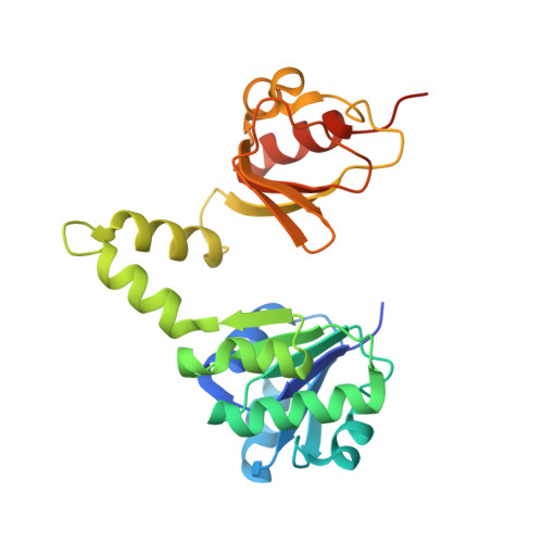
Structural basis of allosteric interactions among Ca(2+)-binding sites in a K(+) channel RCK domain.

Smith, F.J.Pau, V.P.Cingolani, G.Rothberg, B.S.

(2013) Nat Commun 4: 2621-2621

  • DOI: https://doi.org/10.1038/ncomms3621
  • Primary Citation Related Structures: 
    4L73, 4L74, 4L75, 4L76

  • PubMed Abstract: 

    Ligand binding sites within proteins can interact by allosteric mechanisms to modulate binding affinities and control protein function. Here we present crystal structures of the regulator of K+ conductance (RCK) domain from a K+ channel, MthK, which reveal the structural basis of allosteric coupling between two Ca2+ regulatory sites within the domain. Comparison of RCK domain crystal structures in a range of conformations and with different numbers of regulatory Ca2+ ions bound, combined with complementary electrophysiological analysis of channel gating, suggests chemical interactions that are important for modulation of ligand binding and subsequent channel opening.


  • Organizational Affiliation
    • Department of Biochemistry, Temple University School of Medicine, 3400 North Broad Street, Philadelphia, Pennsylvania 19140, USA.

Macromolecule Content 

  • Total Structure Weight: 54.19 kDa 
  • Atom Count: 3,539 
  • Modeled Residue Count: 446 
  • Deposited Residue Count: 484 
  • Unique protein chains: 1

Macromolecules

Find similar proteins by:|  3D Structure
Entity ID: 1
MoleculeChains  Sequence LengthOrganismDetailsImage
Calcium-gated potassium channel MthK
A, B
242Methanothermobacter thermautotrophicus str. Delta HMutation(s): 0 
Gene Names: mthKMTH_1520
Membrane Entity: Yes 
UniProt
Find proteins for O27564 (Methanothermobacter thermautotrophicus (strain ATCC 29096 / DSM 1053 / JCM 10044 / NBRC 100330 / Delta H))
Explore O27564 
Go to UniProtKB:  O27564
Entity Groups
Sequence Clusters30% Identity50% Identity70% Identity90% Identity95% Identity100% Identity
UniProt GroupO27564
Sequence Annotations
Expand
Reference Sequence

Small Molecules

Ligands 3 Unique
IDChains Name / Formula / InChI Key2D Diagram3D Interactions
CA

Query on CA


Download:Ideal Coordinates CCD File
C [auth A],
D [auth A],
E [auth A],
K [auth B],
L [auth B]
CALCIUM ION
Ca
BHPQYMZQTOCNFJ-UHFFFAOYSA-N
CL

Query on CL


Download:Ideal Coordinates CCD File
M [auth B]CHLORIDE ION
Cl
VEXZGXHMUGYJMC-UHFFFAOYSA-M
NA

Query on NA


Download:Ideal Coordinates CCD File
F [auth A]
G [auth A]
H [auth A]
I [auth A]
J [auth A]
F [auth A],
G [auth A],
H [auth A],
I [auth A],
J [auth A],
N [auth B],
O [auth B],
P [auth B]
SODIUM ION
Na
FKNQFGJONOIPTF-UHFFFAOYSA-N

Experimental Data & Validation

Experimental Data

  • Method: X-RAY DIFFRACTION
  • Resolution: 2.50 Å
  • R-Value Free:  0.226 (Depositor), 0.226 (DCC) 
  • R-Value Work:  0.178 (Depositor), 0.177 (DCC) 
  • R-Value Observed: 0.181 (Depositor) 
Space Group: P 31 2 1
Unit Cell:
Length ( Å )Angle ( ˚ )
a = 67.96α = 90
b = 67.96β = 90
c = 159.891γ = 120
Software Package:
Software NamePurpose
SCALAdata scaling
PHASERphasing
PHENIXrefinement
PDB_EXTRACTdata extraction
CBASSdata collection
MOSFLMdata reduction

Structure Validation

View Full Validation Report


Entry History 

Deposition Data

Revision History  (Full details and data files)

  • Version 1.0: 2013-10-23
    Type: Initial release
  • Version 1.1: 2013-11-13
    Changes: Database references
  • Version 1.2: 2023-09-20
    Changes: Data collection, Database references, Derived calculations, Refinement description