4L6Y | pdb_00004l6y

Structure of the microtubule associated protein PRC1 (Protein Regulator of Cytokinesis 1)


Experimental Data Snapshot

  • Method: X-RAY DIFFRACTION
  • Resolution: 3.30 Å
  • R-Value Free: 
    0.306 (Depositor), 0.310 (DCC) 
  • R-Value Work: 
    0.275 (Depositor), 0.275 (DCC) 
  • R-Value Observed: 
    0.276 (Depositor) 

Starting Model: experimental
View more details

wwPDB Validation 3D Report Full Report

Validation slider image for 4L6Y

This is version 1.3 of the entry. See complete history

Literature

Marking and Measuring Single Microtubules by PRC1 and Kinesin-4.

Subramanian, R.Ti, S.C.Tan, L.Darst, S.A.Kapoor, T.M.

(2013) Cell 154: 377-390

  • DOI: https://doi.org/10.1016/j.cell.2013.06.021
  • Primary Citation Related Structures: 
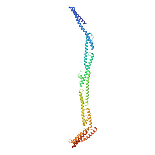
    4L3I, 4L6Y

  • PubMed Abstract: 

    Error-free cell division depends on the assembly of the spindle midzone, a specialized array of overlapping microtubules that emerges between segregating chromosomes during anaphase. The molecular mechanisms by which a subset of dynamic microtubules from the metaphase spindle are selected and organized into a stable midzone array are poorly understood. Here, we show using in vitro reconstitution assays that PRC1 and kinesin-4, two microtubule-associated proteins required for midzone assembly, can tag microtubule plus ends. Remarkably, the size of these tags is proportional to filament length. We determine the crystal structure of the PRC1 homodimer and map the protein-protein interactions needed for tagging microtubule ends. Importantly, length-dependent microtubule plus-end-tagging by PRC1 is also observed in dividing cells. Our findings suggest how biochemically similar microtubules can be differentially marked, based on length, for selective regulation during the formation of specialized arrays, such as those required for cytokinesis.


  • Organizational Affiliation
    • Laboratory of Chemistry and Cell Biology, The Rockefeller University, 1230 York Avenue, New York, NY 10065, USA.

Macromolecule Content 

  • Total Structure Weight: 116.37 kDa 
  • Atom Count: 6,550 
  • Modeled Residue Count: 877 
  • Deposited Residue Count: 980 
  • Unique protein chains: 1

Macromolecules

Find similar proteins by:|  3D Structure
Entity ID: 1
MoleculeChains  Sequence LengthOrganismDetailsImage
Protein regulator of cytokinesis 1
A, B
490Homo sapiensMutation(s): 1 
Gene Names: PRC1
UniProt & NIH Common Fund Data Resources
Find proteins for O43663 (Homo sapiens)
Explore O43663 
Go to UniProtKB:  O43663
PHAROS:  O43663
GTEx:  ENSG00000198901 
Entity Groups
Sequence Clusters30% Identity50% Identity70% Identity90% Identity95% Identity100% Identity
UniProt GroupO43663
Sequence Annotations
Expand
Reference Sequence

Experimental Data & Validation

Experimental Data

  • Method: X-RAY DIFFRACTION
  • Resolution: 3.30 Å
  • R-Value Free:  0.306 (Depositor), 0.310 (DCC) 
  • R-Value Work:  0.275 (Depositor), 0.275 (DCC) 
  • R-Value Observed: 0.276 (Depositor) 
Space Group: P 1 21 1
Unit Cell:
Length ( Å )Angle ( ˚ )
a = 60.66α = 90
b = 209.8β = 105.64
c = 113.41γ = 90
Software Package:
Software NamePurpose
ADSCdata collection
PHASERphasing
PHENIXrefinement
HKL-2000data reduction
HKL-2000data scaling

Structure Validation

View Full Validation Report


Entry History 

Deposition Data

Revision History  (Full details and data files)

  • Version 1.0: 2013-07-17
    Type: Initial release
  • Version 1.1: 2013-08-07
    Changes: Database references
  • Version 1.2: 2017-11-15
    Changes: Refinement description
  • Version 1.3: 2023-09-20
    Changes: Data collection, Database references, Refinement description