4L63 | pdb_00004l63

Apo form of AB5 holotoxin


Experimental Data Snapshot

  • Method: X-RAY DIFFRACTION
  • Resolution: 1.80 Å
  • R-Value Free: 
    0.197 (Depositor), 0.199 (DCC) 
  • R-Value Work: 
    0.177 (Depositor), 0.180 (DCC) 
  • R-Value Observed: 
    0.178 (Depositor) 

Starting Model: experimental
View more details

wwPDB Validation 3D Report Full Report

Validation slider image for 4L63

This is version 1.2 of the entry. See complete history

Literature

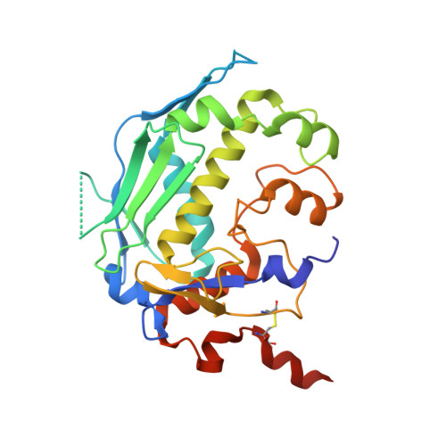
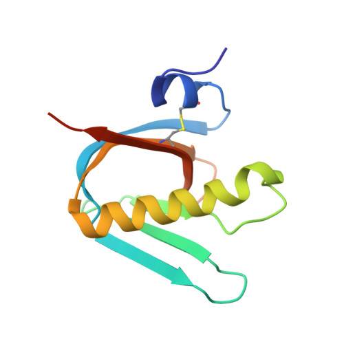
EcxAB Is a Founding Member of a New Family of Metalloprotease AB5 Toxins with a Hybrid Cholera-like B Subunit.

Ng, N.M.Littler, D.R.Paton, A.W.Le Nours, J.Rossjohn, J.Paton, J.C.Beddoe, T.

(2013) Structure 21: 2003-2013

  • DOI: https://doi.org/10.1016/j.str.2013.08.024
  • Primary Citation Related Structures: 
    4L63, 4L6T

  • PubMed Abstract: 

    AB5 toxins are composed of an enzymatic A subunit that disrupts cellular function associated with a pentameric B subunit required for host cell invasion. EcxAB is an AB5 toxin isolated from clinical strains of Escherichia coli classified as part of the cholera family due to B subunit homology. Cholera-group toxins have catalytic ADP-ribosyltransferases as their A subunits, so it was surprising that EcxA did not. We confirmed that EcxAB self-associates as a functional toxin and obtained its structure. EcxAB is a prototypical member of a hybrid AB5 toxin family containing metzincin-type metalloproteases as their active A subunit paired to a cholera-like B subunit. Furthermore, EcxA is distinct from previously characterized proteases and thus founds an AB5-associated metzincin family that we term the toxilysins. EcxAB provides the first observation of conserved B subunit usage across different AB5 toxin families and provides evidence that the intersubunit interface of these toxins is far more permissive than previously supposed.


  • Organizational Affiliation
    • ARC Centre of Excellence in Structural and Functional Microbial Genomics, Department of Biochemistry and Molecular Biology, School of Biomedical Sciences, Monash University, Clayton, VIC 3800, Australia.

Macromolecule Content 

  • Total Structure Weight: 94.63 kDa 
  • Atom Count: 6,569 
  • Modeled Residue Count: 771 
  • Deposited Residue Count: 826 
  • Unique protein chains: 2

Macromolecules

Find similar proteins by:|  3D Structure
Entity ID: 1
MoleculeChains  Sequence LengthOrganismDetailsImage
ECXA266Escherichia coliMutation(s): 0 
Gene Names: ecxA
EC: 3.4.24
UniProt
Find proteins for Q8GAV4 (Escherichia coli)
Explore Q8GAV4 
Go to UniProtKB:  Q8GAV4
Entity Groups
Sequence Clusters30% Identity50% Identity70% Identity90% Identity95% Identity100% Identity
UniProt GroupQ8GAV4
Sequence Annotations
Expand
Reference Sequence
Find similar proteins by:|  3D Structure
Entity ID: 2
MoleculeChains  Sequence LengthOrganismDetailsImage
ECXB
B, C, D, E, F
112Escherichia coliMutation(s): 0 
Gene Names: ecxB
UniProt
Find proteins for Q8GAV3 (Escherichia coli)
Explore Q8GAV3 
Go to UniProtKB:  Q8GAV3
Entity Groups
Sequence Clusters30% Identity50% Identity70% Identity90% Identity95% Identity100% Identity
UniProt GroupQ8GAV3
Sequence Annotations
Expand
Reference Sequence

Experimental Data & Validation

Experimental Data

  • Method: X-RAY DIFFRACTION
  • Resolution: 1.80 Å
  • R-Value Free:  0.197 (Depositor), 0.199 (DCC) 
  • R-Value Work:  0.177 (Depositor), 0.180 (DCC) 
  • R-Value Observed: 0.178 (Depositor) 
Space Group: P 65 2 2
Unit Cell:
Length ( Å )Angle ( ˚ )
a = 120.53α = 90
b = 120.53β = 90
c = 273.51γ = 120
Software Package:
Software NamePurpose
ADSCdata collection
PHASERphasing
PHENIXrefinement
MOSFLMdata reduction
SCALAdata scaling

Structure Validation

View Full Validation Report


Entry History 

Deposition Data

Revision History  (Full details and data files)

  • Version 1.0: 2013-11-27
    Type: Initial release
  • Version 1.1: 2023-09-20
    Changes: Data collection, Database references, Derived calculations, Refinement description
  • Version 1.2: 2024-11-06
    Changes: Structure summary