4L5Z | pdb_00004l5z

Crystal structure of the Candida albicans Methionine Synthase in complex with Homocysteine


Experimental Data Snapshot

  • Method: X-RAY DIFFRACTION
  • Resolution: 2.18 Å
  • R-Value Free: 
    0.235 (Depositor), 0.226 (DCC) 
  • R-Value Work: 
    0.173 (Depositor) 
  • R-Value Observed: 
    0.176 (Depositor) 

wwPDB Validation 3D Report Full Report

Validation slider image for 4L5Z

This is version 1.3 of the entry. See complete history

Literature

Structural analysis of a fungal methionine synthase with substrates and inhibitors.

Ubhi, D.Kago, G.Monzingo, A.F.Robertus, J.D.

(2014) J Mol Biology 426: 1839-1847

  • DOI: https://doi.org/10.1016/j.jmb.2014.02.006
  • Primary Citation Related Structures: 
    4L5Z, 4L61, 4L64, 4L65, 4L6H, 4L6O

  • PubMed Abstract: 

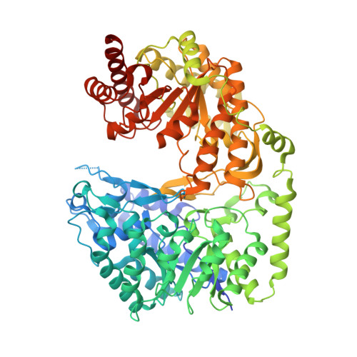
    The cobalamin-independent methionine synthase from Candida albicans, known as Met6p, is a 90-kDa enzyme that consists of two (βα)8 barrels. The active site is located between the two domains and has binding sites for a zinc ion and substrates L-homocysteine and 5-methyl-tetrahydrofolate-glutamate3. Met6p catalyzes transfer of the methyl group of 5-methyl-tetrahydrofolate-glutamate3 to the L-homocysteine thiolate to generate methionine. Met6p is essential for fungal growth, and we currently pursue it as an antifungal drug design target. Here we report the binding of L-homocysteine, methionine, and several folate analogs. We show that binding of L-homocysteine or methionine results in conformational rearrangements at the amino acid binding pocket, moving the catalytic zinc into position to activate the thiol group. We also map the folate binding pocket and identify specific binding residues, like Asn126, whose mutation eliminates catalytic activity. We also report the development of a robust fluorescence-based activity assay suitable for high-throughput screening. We use this assay and an X-ray structure to characterize methotrexate as a weak inhibitor of fungal Met6p.


  • Organizational Affiliation
    • Institute for Cellular and Molecular Biology, Department of Molecular Biosciences, University of Texas at Austin, Austin, TX 78712, USA.

Macromolecule Content 

  • Total Structure Weight: 88.39 kDa 
  • Atom Count: 6,088 
  • Modeled Residue Count: 753 
  • Deposited Residue Count: 789 
  • Unique protein chains: 1

Macromolecules

Find similar proteins by:|  3D Structure
Entity ID: 1
MoleculeChains  Sequence LengthOrganismDetailsImage
5-methyltetrahydropteroyltriglutamate--homocysteine methyltransferase789Candida albicans SC5314Mutation(s): 3 
Gene Names: CaO19.10083CaO19.2551MET6
EC: 2.1.1.14
UniProt
Find proteins for P82610 (Candida albicans (strain SC5314 / ATCC MYA-2876))
Explore P82610 
Go to UniProtKB:  P82610
Entity Groups
Sequence Clusters30% Identity50% Identity70% Identity90% Identity95% Identity100% Identity
UniProt GroupP82610
Sequence Annotations
Expand
Reference Sequence

Experimental Data & Validation

Experimental Data

  • Method: X-RAY DIFFRACTION
  • Resolution: 2.18 Å
  • R-Value Free:  0.235 (Depositor), 0.226 (DCC) 
  • R-Value Work:  0.173 (Depositor) 
  • R-Value Observed: 0.176 (Depositor) 
Space Group: P 21 21 21
Unit Cell:
Length ( Å )Angle ( ˚ )
a = 77.124α = 90
b = 97.748β = 90
c = 98.194γ = 90
Software Package:
Software NamePurpose
ADSCdata collection
PHASERphasing
REFMACrefinement
HKL-2000data reduction
HKL-2000data scaling

Structure Validation

View Full Validation Report


Entry History 

Deposition Data

Revision History  (Full details and data files)

  • Version 1.0: 2014-03-05
    Type: Initial release
  • Version 1.1: 2014-04-09
    Changes: Database references
  • Version 1.2: 2024-02-28
    Changes: Data collection, Database references, Derived calculations
  • Version 1.3: 2024-04-03
    Changes: Structure summary