4L5N

Crystallographic Structure of HHV-1 Uracil-DNA Glycosylase complexed with the Bacillus phage PZA inhibitor protein p56


Experimental Data Snapshot

  • Method: X-RAY DIFFRACTION
  • Resolution: 2.16 Å
  • R-Value Free: 0.202 
  • R-Value Work: 0.171 
  • R-Value Observed: 0.172 

wwPDB Validation   3D Report Full Report


This is version 1.3 of the entry. See complete history


Literature

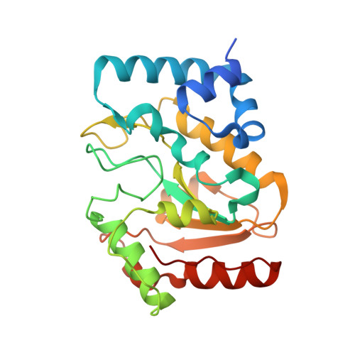
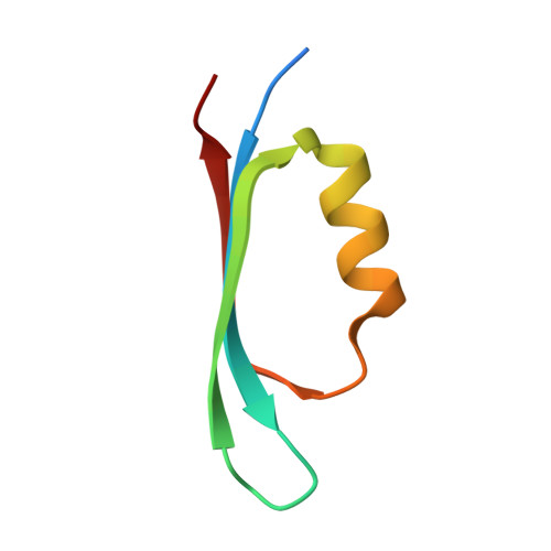
Architecturally diverse proteins converge on an analogous mechanism to inactivate Uracil-DNA glycosylase.

Cole, A.R.Ofer, S.Ryzhenkova, K.Baltulionis, G.Hornyak, P.Savva, R.

(2013) Nucleic Acids Res 41: 8760-8775

  • DOI: https://doi.org/10.1093/nar/gkt633
  • Primary Citation of Related Structures:  
    4L5N

  • PubMed Abstract: 

    Uracil-DNA glycosylase (UDG) compromises the replication strategies of diverse viruses from unrelated lineages. Virally encoded proteins therefore exist to limit, inhibit or target UDG activity for proteolysis. Viral proteins targeting UDG, such as the bacteriophage proteins ugi, and p56, and the HIV-1 protein Vpr, share no sequence similarity, and are not structurally homologous. Such diversity has hindered identification of known or expected UDG-inhibitory activities in other genomes. The structural basis for UDG inhibition by ugi is well characterized; yet, paradoxically, the structure of the unbound p56 protein is enigmatically unrevealing of its mechanism. To resolve this conundrum, we determined the structure of a p56 dimer bound to UDG. A helix from one of the subunits of p56 occupies the UDG DNA-binding cleft, whereas the dimer interface forms a hydrophobic box to trap a mechanistically important UDG residue. Surprisingly, these p56 inhibitory elements are unexpectedly analogous to features used by ugi despite profound architectural disparity. Contacts from B-DNA to UDG are mimicked by residues of the p56 helix, echoing the role of ugi's inhibitory beta strand. Using mutagenesis, we propose that DNA mimicry by p56 is a targeting and specificity mechanism supporting tight inhibition via hydrophobic sequestration.


  • Organizational Affiliation

    Department of Biological Sciences, Institute of Structural and Molecular Biology, Birkbeck College, University of London, Malet Street, London WC1E 7HX, UK and Research Department of Structural and Molecular Biology, Institute of Structural and Molecular Biology, University College London, Gower Street, London WC1E 6BT, UK.


Macromolecules
Find similar proteins by:  (by identity cutoff)  |  3D Structure
Entity ID: 1
MoleculeChains Sequence LengthOrganismDetailsImage
Uracil-DNA glycosylase
A, B
241Human alphaherpesvirus 1 strain 17Mutation(s): 0 
Gene Names: UL2
EC: 3.2.2.27
UniProt
Find proteins for P10186 (Human herpesvirus 1 (strain 17))
Explore P10186 
Go to UniProtKB:  P10186
Entity Groups  
Sequence Clusters30% Identity50% Identity70% Identity90% Identity95% Identity100% Identity
UniProt GroupP10186
Sequence Annotations
Expand
  • Reference Sequence
Find similar proteins by:  (by identity cutoff)  |  3D Structure
Entity ID: 2
MoleculeChains Sequence LengthOrganismDetailsImage
Early protein GP1B
C, D, E, F
55Bacillus phage PZAMutation(s): 0 
Gene Names: 1B
UniProt
Find proteins for P06948 (Bacillus phage PZA)
Explore P06948 
Go to UniProtKB:  P06948
Entity Groups  
Sequence Clusters30% Identity50% Identity70% Identity90% Identity95% Identity100% Identity
UniProt GroupP06948
Sequence Annotations
Expand
  • Reference Sequence
Small Molecules
Ligands 1 Unique
IDChains Name / Formula / InChI Key2D Diagram3D Interactions
ACT
Query on ACT

Download Ideal Coordinates CCD File 
G [auth A]
H [auth A]
I [auth A]
J [auth A]
K [auth A]
G [auth A],
H [auth A],
I [auth A],
J [auth A],
K [auth A],
L [auth A],
M [auth A],
N [auth B],
O [auth B],
P [auth C],
Q [auth D],
R [auth E],
S [auth F],
T [auth F],
U [auth F]
ACETATE ION
C2 H3 O2
QTBSBXVTEAMEQO-UHFFFAOYSA-M
Experimental Data & Validation

Experimental Data

  • Method: X-RAY DIFFRACTION
  • Resolution: 2.16 Å
  • R-Value Free: 0.202 
  • R-Value Work: 0.171 
  • R-Value Observed: 0.172 
  • Space Group: P 21 21 21
Unit Cell:
Length ( Å )Angle ( ˚ )
a = 52.704α = 90
b = 91.375β = 90
c = 162.862γ = 90
Software Package:
Software NamePurpose
GDAdata collection
PHASERphasing
BUSTERrefinement
XDSdata reduction
Aimlessdata scaling

Structure Validation

View Full Validation Report


Entry History 

Deposition Data

Revision History  (Full details and data files)

  • Version 1.0: 2013-08-07
    Type: Initial release
  • Version 1.1: 2013-08-21
    Changes: Database references
  • Version 1.2: 2013-10-23
    Changes: Database references
  • Version 1.3: 2024-02-28
    Changes: Data collection, Database references, Derived calculations