4L3E

The complex between high affinity TCR DMF5(alpha-D26Y,beta-L98W) and human Class I MHC HLA-A2 with the bound MART-1(26-35)(A27L) peptide


Experimental Data Snapshot

  • Method: X-RAY DIFFRACTION
  • Resolution: 2.56 Å
  • R-Value Free: 0.266 
  • R-Value Work: 0.229 
  • R-Value Observed: 0.231 

wwPDB Validation   3D Report Full Report


This is version 1.1 of the entry. See complete history


Literature

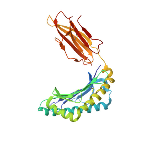
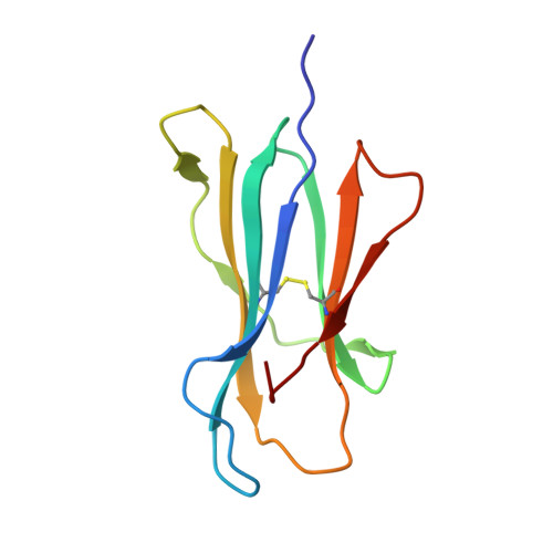
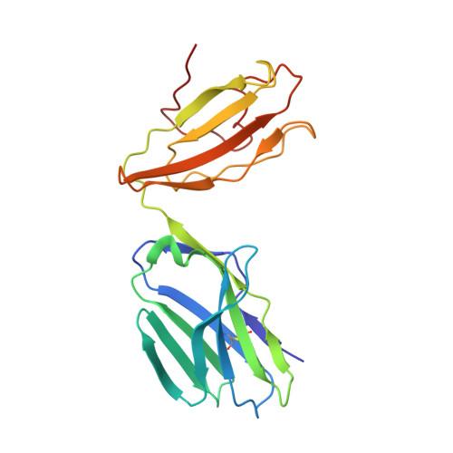
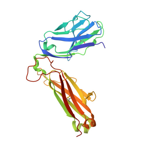
Computational design of the affinity and specificity of a therapeutic T cell receptor.

Pierce, B.G.Hellman, L.M.Hossain, M.Singh, N.K.Vander Kooi, C.W.Weng, Z.Baker, B.M.

(2014) PLoS Comput Biol 10: e1003478-e1003478

  • DOI: https://doi.org/10.1371/journal.pcbi.1003478
  • Primary Citation of Related Structures:  
    4L3E

  • PubMed Abstract: 

    T cell receptors (TCRs) are key to antigen-specific immunity and are increasingly being explored as therapeutics, most visibly in cancer immunotherapy. As TCRs typically possess only low-to-moderate affinity for their peptide/MHC (pMHC) ligands, there is a recognized need to develop affinity-enhanced TCR variants. Previous in vitro engineering efforts have yielded remarkable improvements in TCR affinity, yet concerns exist about the maintenance of peptide specificity and the biological impacts of ultra-high affinity. As opposed to in vitro engineering, computational design can directly address these issues, in theory permitting the rational control of peptide specificity together with relatively controlled increments in affinity. Here we explored the efficacy of computational design with the clinically relevant TCR DMF5, which recognizes nonameric and decameric epitopes from the melanoma-associated Melan-A/MART-1 protein presented by the class I MHC HLA-A2. We tested multiple mutations selected by flexible and rigid modeling protocols, assessed impacts on affinity and specificity, and utilized the data to examine and improve algorithmic performance. We identified multiple mutations that improved binding affinity, and characterized the structure, affinity, and binding kinetics of a previously reported double mutant that exhibits an impressive 400-fold affinity improvement for the decameric pMHC ligand without detectable binding to non-cognate ligands. The structure of this high affinity mutant indicated very little conformational consequences and emphasized the high fidelity of our modeling procedure. Overall, our work showcases the capability of computational design to generate TCRs with improved pMHC affinities while explicitly accounting for peptide specificity, as well as its potential for generating TCRs with customized antigen targeting capabilities.


  • Organizational Affiliation

    Program in Bioinformatics and Integrative Biology, University of Massachusetts Medical School, Worcester, Massachusetts, United States of America.


Macromolecules
Find similar proteins by:  (by identity cutoff)  |  3D Structure
Entity ID: 1
MoleculeChains Sequence LengthOrganismDetailsImage
HLA class I histocompatibility antigen, A-2 alpha chain275Homo sapiensMutation(s): 0 
Gene Names: HLA-AHLAA
UniProt & NIH Common Fund Data Resources
Find proteins for P04439 (Homo sapiens)
Explore P04439 
Go to UniProtKB:  P04439
PHAROS:  P04439
GTEx:  ENSG00000206503 
Entity Groups  
Sequence Clusters30% Identity50% Identity70% Identity90% Identity95% Identity100% Identity
UniProt GroupP04439
Sequence Annotations
Expand
  • Reference Sequence
Find similar proteins by:  (by identity cutoff)  |  3D Structure
Entity ID: 2
MoleculeChains Sequence LengthOrganismDetailsImage
Beta-2-microglobulin100Homo sapiensMutation(s): 0 
Gene Names: B2MCDABP0092HDCMA22P
UniProt & NIH Common Fund Data Resources
Find proteins for P61769 (Homo sapiens)
Explore P61769 
Go to UniProtKB:  P61769
PHAROS:  P61769
GTEx:  ENSG00000166710 
Entity Groups  
Sequence Clusters30% Identity50% Identity70% Identity90% Identity95% Identity100% Identity
UniProt GroupP61769
Sequence Annotations
Expand
  • Reference Sequence

Find similar proteins by:  Sequence   |   3D Structure  

Entity ID: 3
MoleculeChains Sequence LengthOrganismDetailsImage
Melanoma antigen recognized by T-cells 110Homo sapiensMutation(s): 1 
UniProt & NIH Common Fund Data Resources
Find proteins for Q16655 (Homo sapiens)
Explore Q16655 
Go to UniProtKB:  Q16655
PHAROS:  Q16655
GTEx:  ENSG00000120215 
Entity Groups  
Sequence Clusters30% Identity50% Identity70% Identity90% Identity95% Identity100% Identity
UniProt GroupQ16655
Sequence Annotations
Expand
  • Reference Sequence
Find similar proteins by:  (by identity cutoff)  |  3D Structure
Entity ID: 4
MoleculeChains Sequence LengthOrganismDetailsImage
DMF5 alpha chain199Homo sapiensMutation(s): 0 
Gene Names: TRACTCRA
Entity Groups  
Sequence Clusters30% Identity50% Identity70% Identity90% Identity95% Identity100% Identity
Sequence Annotations
Expand
  • Reference Sequence
Find similar proteins by:  (by identity cutoff)  |  3D Structure
Entity ID: 5
MoleculeChains Sequence LengthOrganismDetailsImage
DMF5 beta chain242Homo sapiensMutation(s): 0 
Entity Groups  
Sequence Clusters30% Identity50% Identity70% Identity90% Identity95% Identity100% Identity
Sequence Annotations
Expand
  • Reference Sequence
Experimental Data & Validation

Experimental Data

  • Method: X-RAY DIFFRACTION
  • Resolution: 2.56 Å
  • R-Value Free: 0.266 
  • R-Value Work: 0.229 
  • R-Value Observed: 0.231 
  • Space Group: C 1 2 1
Unit Cell:
Length ( Å )Angle ( ˚ )
a = 227.04α = 90
b = 49.316β = 94.82
c = 92.894γ = 90
Software Package:
Software NamePurpose
HKL-2000data collection
PHENIXmodel building
PHENIXrefinement
HKL-2000data reduction
HKL-2000data scaling
PHENIXphasing

Structure Validation

View Full Validation Report


Entry History 

Deposition Data

Revision History  (Full details and data files)

  • Version 1.0: 2014-06-11
    Type: Initial release
  • Version 1.1: 2014-08-27
    Changes: Database references