4L2M

Crystal structure of the 2/2 hemoglobin from Synechococcus sp. PCC 7002 in the cyanomet state and with covalently attached heme


Experimental Data Snapshot

  • Method: X-RAY DIFFRACTION
  • Resolution: 2.25 Å
  • R-Value Free: 0.288 
  • R-Value Work: 0.218 
  • R-Value Observed: 0.221 

wwPDB Validation   3D Report Full Report


Ligand Structure Quality Assessment 


This is version 1.4 of the entry. See complete history


Literature

The 2/2 hemoglobin from the cyanobacterium Synechococcus sp. PCC 7002 with covalently attached heme: Comparison of X-ray and NMR structures.

Wenke, B.B.Lecomte, J.T.Heroux, A.Schlessman, J.L.

(2014) Proteins 82: 528-534

  • DOI: https://doi.org/10.1002/prot.24409
  • Primary Citation of Related Structures:  
    4L2M, 4MAX

  • PubMed Abstract: 

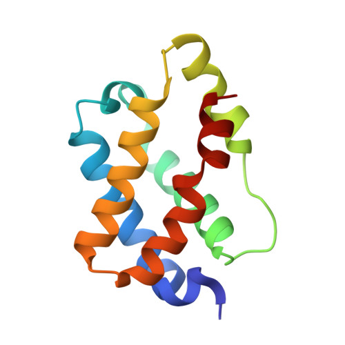
    The X-ray structures of the hemoglobin from Synechococcus sp. PCC 7002 (GlbN) were solved in the ferric bis-histidine (1.44 Å resolution) and cyanide-bound (2.25 Å resolution) states with covalently attached heme. The two structures illustrate the conformational changes and cavity opening caused by exogenous ligand binding. They also reveal an unusually distorted heme, ruffled as in c cytochromes. Comparison to the solution structure demonstrates the influence of crystal packing on several structural elements, whereas comparison to GlbN from Synechocystis sp. PCC 6803 shows subtle differences in heme geometries and environment. The new structures will be instrumental in elucidating GlbN reactivity.


  • Organizational Affiliation

    T.C. Jenkins Department of Biophysics, Johns Hopkins University, Baltimore, Maryland, 21218.


Macromolecules
Find similar proteins by:  (by identity cutoff)  |  3D Structure
Entity ID: 1
MoleculeChains Sequence LengthOrganismDetailsImage
Cyanoglobin
A, B
123Picosynechococcus sp. PCC 7002Mutation(s): 0 
Gene Names: glbNSYNPCC7002_A1621
UniProt
Find proteins for Q8RT58 (Picosynechococcus sp. (strain ATCC 27264 / PCC 7002 / PR-6))
Explore Q8RT58 
Go to UniProtKB:  Q8RT58
Entity Groups  
Sequence Clusters30% Identity50% Identity70% Identity90% Identity95% Identity100% Identity
UniProt GroupQ8RT58
Sequence Annotations
Expand
  • Reference Sequence
Experimental Data & Validation

Experimental Data

  • Method: X-RAY DIFFRACTION
  • Resolution: 2.25 Å
  • R-Value Free: 0.288 
  • R-Value Work: 0.218 
  • R-Value Observed: 0.221 
  • Space Group: P 1 21 1
Unit Cell:
Length ( Å )Angle ( ˚ )
a = 50.622α = 90
b = 40.951β = 111.21
c = 66.461γ = 90
Software Package:
Software NamePurpose
SCALEPACKdata scaling
PHASERphasing
REFMACrefinement
PDB_EXTRACTdata extraction
CBASSdata collection
HKL-2000data reduction

Structure Validation

View Full Validation Report


Ligand Structure Quality Assessment 


Entry History 

Deposition Data

Revision History  (Full details and data files)

  • Version 1.0: 2013-06-12
    Type: Initial release
  • Version 1.1: 2013-06-19
    Changes: Experimental preparation
  • Version 1.2: 2013-09-18
    Changes: Database references
  • Version 1.3: 2014-02-19
    Changes: Database references
  • Version 1.4: 2023-09-20
    Changes: Data collection, Database references, Derived calculations, Refinement description