4L2H | pdb_00004l2h

Structure of a catalytically inactive PARG in complex with a poly-ADP-ribose fragment


Experimental Data Snapshot

  • Method: X-RAY DIFFRACTION
  • Resolution: 1.46 Å
  • R-Value Free: 
    0.177 (Depositor), 0.180 (DCC) 
  • R-Value Work: 
    0.138 (Depositor), 0.147 (DCC) 
  • R-Value Observed: 
    0.140 (Depositor) 

Starting Model: experimental
View more details

wwPDB Validation 3D Report Full Report

Validation slider image for 4L2H

Ligand Structure Quality Assessment 


This is version 1.3 of the entry. See complete history

Literature

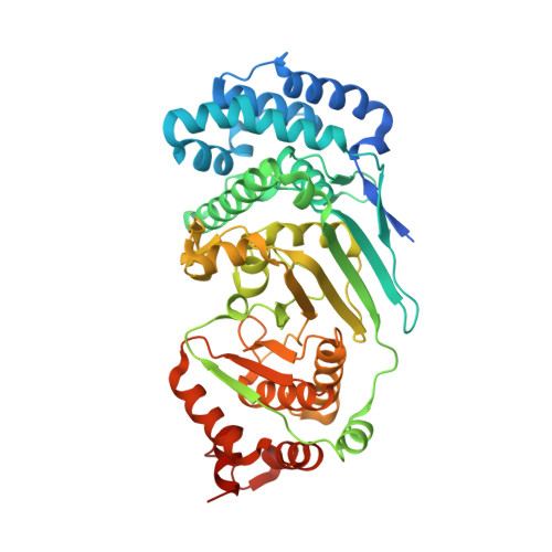
Visualization of poly(ADP-ribose) bound to PARG reveals inherent balance between exo- and endo-glycohydrolase activities.

Barkauskaite, E.Brassington, A.Tan, E.S.Warwicker, J.Dunstan, M.S.Banos, B.Lafite, P.Ahel, M.Mitchison, T.J.Ahel, I.Leys, D.

(2013) Nat Commun 4: 2164-2164

  • DOI: https://doi.org/10.1038/ncomms3164
  • Primary Citation Related Structures: 
    4L2H

  • PubMed Abstract: 

    Poly-ADP-ribosylation is a post-translational modification that regulates processes involved in genome stability. Breakdown of the poly(ADP-ribose) (PAR) polymer is catalysed by poly(ADP-ribose) glycohydrolase (PARG), whose endo-glycohydrolase activity generates PAR fragments. Here we present the crystal structure of PARG incorporating the PAR substrate. The two terminal ADP-ribose units of the polymeric substrate are bound in exo-mode. Biochemical and modelling studies reveal that PARG acts predominantly as an exo-glycohydrolase. This preference is linked to Phe902 (human numbering), which is responsible for low-affinity binding of the substrate in endo-mode. Our data reveal the mechanism of poly-ADP-ribosylation reversal, with ADP-ribose as the dominant product, and suggest that the release of apoptotic PAR fragments occurs at unusual PAR/PARG ratios.


  • Organizational Affiliation
    • Cancer Research UK, Paterson Institute for Cancer Research, University of Manchester, Wilmslow Road, Manchester M20 4BX, UK.

Macromolecule Content 

  • Total Structure Weight: 57 kDa 
  • Atom Count: 4,186 
  • Modeled Residue Count: 436 
  • Deposited Residue Count: 477 
  • Unique protein chains: 1

Macromolecules

Find similar proteins by:|  3D Structure
Entity ID: 1
MoleculeChains  Sequence LengthOrganismDetailsImage
Poly(ADP-ribose) glycohydrolase477Tetrahymena thermophilaMutation(s): 1 
EC: 3.2.1.143 (PDB Primary Data), 3.2.2 (UniProt)
UniProt
Find proteins for I6L8L7 (Tetrahymena thermophila)
Explore I6L8L7 
Go to UniProtKB:  I6L8L7
Entity Groups
Sequence Clusters30% Identity50% Identity70% Identity90% Identity95% Identity100% Identity
UniProt GroupI6L8L7
Sequence Annotations
Expand
Reference Sequence

Small Molecules

Ligands 1 Unique
IDChains Name / Formula / InChI Key2D Diagram3D Interactions
AR6

Query on AR6


Download:Ideal Coordinates CCD File
B [auth A],
C [auth A]
[(2R,3S,4R,5R)-5-(6-AMINOPURIN-9-YL)-3,4-DIHYDROXY-OXOLAN-2-YL]METHYL[HYDROXY-[[(2R,3S,4R,5S)-3,4,5-TRIHYDROXYOXOLAN-2-YL]METHOXY]PHOSPHORYL] HYDROGEN PHOSPHATE
C15 H23 N5 O14 P2
SRNWOUGRCWSEMX-ZQSHOCFMSA-N

Experimental Data & Validation

Experimental Data

  • Method: X-RAY DIFFRACTION
  • Resolution: 1.46 Å
  • R-Value Free:  0.177 (Depositor), 0.180 (DCC) 
  • R-Value Work:  0.138 (Depositor), 0.147 (DCC) 
  • R-Value Observed: 0.140 (Depositor) 
Space Group: P 21 21 21
Unit Cell:
Length ( Å )Angle ( ˚ )
a = 55.928α = 90
b = 75.713β = 90
c = 138.957γ = 90
Software Package:
Software NamePurpose
PHASERphasing
REFMACrefinement
XDSdata reduction
XDSdata scaling

Structure Validation

View Full Validation Report


Ligand Structure Quality Assessment 


Entry History 

Deposition Data

Revision History  (Full details and data files)

  • Version 1.0: 2013-07-24
    Type: Initial release
  • Version 1.1: 2013-08-07
    Changes: Database references
  • Version 1.2: 2013-08-21
    Changes: Database references
  • Version 1.3: 2023-09-20
    Changes: Data collection, Database references, Derived calculations, Refinement description, Structure summary