Crystal Structure of Metallo DNA Duplex Containing Consecutive Watson-Crick-like T-Hg(II) -T Base Pairs
Kondo, J., Yamada, T., Hirose, C., Okamoto, I., Tanaka, Y., Ono, A.(2014) Angew Chem Int Ed Engl 53: 2385-2388
- PubMed: 24478025 Search on PubMed
- DOI: https://doi.org/10.1002/anie.201309066
- Primary Citation Related Structures:
4L24, 4L25, 4L26 - PubMed Abstract:
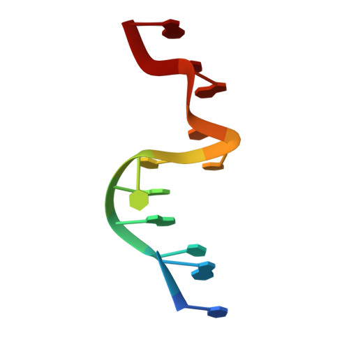
The metallo DNA duplex containing mercury-mediated T-T base pairs is an attractive biomacromolecular nanomaterial which can be applied to nanodevices such as ion sensors. Reported herein is the first crystal structure of a B-form DNA duplex containing two consecutive T-Hg(II)-T base pairs. The Hg(II) ion occupies the center between two T residues. The N3-Hg(II) bond distance is 2.0 Å. The relatively short Hg(II)-Hg(II) distance (3.3 Å) observed in consecutive T-Hg(II)-T base pairs suggests that the metallophilic attraction could exist between them and may stabilize the B-form double helix. To support this, the DNA duplex is largely distorted and adopts an unusual nonhelical conformation in the absence of Hg(II). The structure of the metallo DNA duplex itself and the Hg(II)-induced structural switching from the nonhelical form to the B-form provide the basis for structure-based design of metal-conjugated nucleic acid nanomaterials.
- Department of Materials and Life Sciences, Faculty of Science and Technology, Sophia University, 7-1 Kioi-cho, Chiyoda-ku, 102-8554 Tokyo (Japan). j.kondo@sophia.ac.jp.
Organizational Affiliation: