Cd(2+) as a ca(2+) surrogate in protein-membrane interactions: isostructural but not isofunctional.
Morales, K.A., Yang, Y., Long, Z., Li, P., Taylor, A.B., Hart, P.J., Igumenova, T.I.(2013) J Am Chem Soc 135: 12980-12983
- PubMed: 23937054 Search on PubMedSearch on PubMed Central
- DOI: https://doi.org/10.1021/ja406958k
- Primary Citation Related Structures:
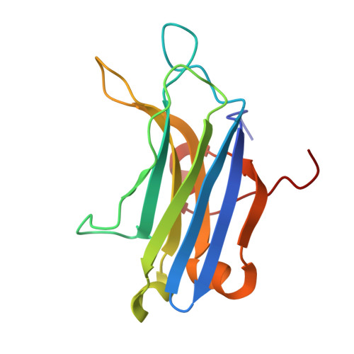
4L1L - PubMed Abstract:
Due to its favorable spectroscopic properties, Cd(2+) is frequently used as a probe of Ca(2+) sites in proteins. We investigate the ability of Cd(2+) to act as a structural and functional surrogate of Ca(2+) in protein-membrane interactions. C2 domain from protein kinase Cα (C2α) was chosen as a paradigm for the Ca(2+)-dependent phosphatidylserine-binding peripheral membrane domains. We identified the Cd(2+)-binding sites of C2α using NMR spectroscopy, determined the 1.6 Å crystal structure of Cd(2+)-bound C2α, and characterized metal-ion-dependent interactions between C2α and phospholipid membranes using fluorescence spectroscopy and ultracentrifugation experiments. We show that Cd(2+) forms a tight complex with the membrane-binding loops of C2α but is unable to support its membrane-binding function. This is in sharp contrast with Pb(2+), which is almost as effective as Ca(2+) in driving the C2α-membrane association process. Our results provide the first direct evidence for the specific role of divalent metal ions in mediating protein-membrane interactions, have important implications for metal substitution studies in proteins, and illustrate the potential diversity of functional responses caused by toxic metal ions.
- Department of Biochemistry and Biophysics, Texas A&M University, College Station, Texas 77843, United States.
Organizational Affiliation: