4L1K

Crystal structure of D-alanine-D-alnine ligase from Xanthomonas oryzae pv. oryzae with AMPPNP


Experimental Data Snapshot

  • Method: X-RAY DIFFRACTION
  • Resolution: 2.30 Å
  • R-Value Free: 0.264 
  • R-Value Work: 0.211 
  • R-Value Observed: 0.215 

wwPDB Validation   3D Report Full Report


Ligand Structure Quality Assessment 


This is version 1.2 of the entry. See complete history


Literature

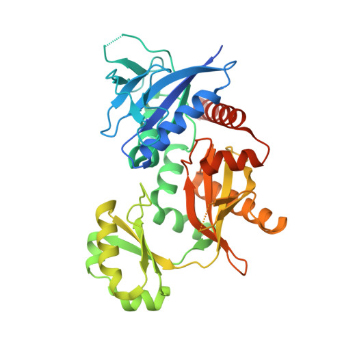
Crystal structures of d-alanine-d-alanine ligase from Xanthomonas oryzae pv. oryzae alone and in complex with nucleotides

Doan, T.T.N.Kim, J.K.Ngo, H.P.T.Tran, H.T.Cha, S.S.Chung, K.M.Huynh, K.H.Ahn, Y.J.Kang, L.W.

(2014) Arch Biochem Biophys 545C: 92-99

  • DOI: https://doi.org/10.1016/j.abb.2014.01.009
  • Primary Citation of Related Structures:  
    4L1K, 4ME6

  • PubMed Abstract: 

    D-Alanine-D-alanine ligase (DDL) catalyzes the biosynthesis of d-alanyl-d-alanine, an essential bacterial peptidoglycan precursor, and is an important drug target for the development of antibacterials. We determined four different crystal structures of DDL from Xanthomonas oryzae pv. oryzae (Xoo) causing Bacteria Blight (BB), which include apo, ADP-bound, ATP-bound, and AMPPNP-bound structures at the resolution between 2.3 and 2.0 Å. Similarly with other DDLs, the active site of XoDDL is formed by three loops from three domains at the center of enzyme. Compared with d-alanyl-d-alanine and ATP-bound TtDDL structure, the γ-phosphate of ATP in XoDDL structure was shifted outside toward solution. We swapped the ω-loop (loop3) of XoDDL with those of Escherichia coli and Helicobacter pylori DDLs, and measured the enzymatic kinetics of wild-type XoDDL and two mutant XoDDLs with the swapped ω-loops. Results showed that the direct interactions between ω-loop and other two loops are essential for the active ATP conformation for D-ala-phosphate formation.


  • Organizational Affiliation

    Department of Advanced Technology Fusion, Konkuk University, Hwayang dong, Gwangjin-gu, Seoul 143-01, Republic of Korea.


Macromolecules
Find similar proteins by:  (by identity cutoff)  |  3D Structure
Entity ID: 1
MoleculeChains Sequence LengthOrganismDetailsImage
D-alanine--D-alanine ligase384Xanthomonas oryzae pv. oryzae KACC 10331Mutation(s): 1 
Gene Names: ddlddlAXOO0352
EC: 6.3.2.4
UniProt
Find proteins for Q5H614 (Xanthomonas oryzae pv. oryzae (strain KACC10331 / KXO85))
Explore Q5H614 
Go to UniProtKB:  Q5H614
Entity Groups  
Sequence Clusters30% Identity50% Identity70% Identity90% Identity95% Identity100% Identity
UniProt GroupQ5H614
Sequence Annotations
Expand
  • Reference Sequence
Small Molecules
Ligands 2 Unique
IDChains Name / Formula / InChI Key2D Diagram3D Interactions
ANP
Query on ANP

Download Ideal Coordinates CCD File 
B [auth A]PHOSPHOAMINOPHOSPHONIC ACID-ADENYLATE ESTER
C10 H17 N6 O12 P3
PVKSNHVPLWYQGJ-KQYNXXCUSA-N
MG
Query on MG

Download Ideal Coordinates CCD File 
C [auth A]MAGNESIUM ION
Mg
JLVVSXFLKOJNIY-UHFFFAOYSA-N
Experimental Data & Validation

Experimental Data

  • Method: X-RAY DIFFRACTION
  • Resolution: 2.30 Å
  • R-Value Free: 0.264 
  • R-Value Work: 0.211 
  • R-Value Observed: 0.215 
  • Space Group: P 43 21 2
Unit Cell:
Length ( Å )Angle ( ˚ )
a = 83.018α = 90
b = 83.018β = 90
c = 97.364γ = 90
Software Package:
Software NamePurpose
HKL-2000data collection
MOLREPphasing
REFMACrefinement
HKL-2000data reduction
SCALEPACKdata scaling

Structure Validation

View Full Validation Report


Ligand Structure Quality Assessment 


Entry History 

Deposition Data

Revision History  (Full details and data files)

  • Version 1.0: 2014-02-19
    Type: Initial release
  • Version 1.1: 2017-11-15
    Changes: Refinement description
  • Version 1.2: 2024-03-20
    Changes: Data collection, Database references, Derived calculations