4L1J | pdb_00004l1j

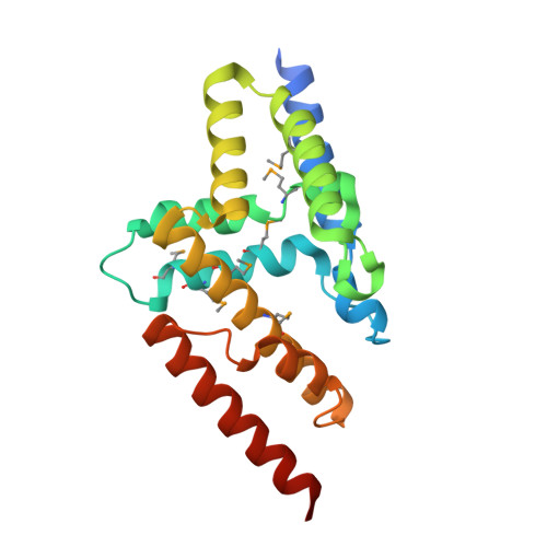
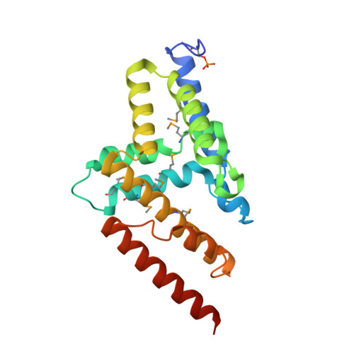
Three dimensional structure of mutant D143A of human HD domain-containing protein 2, Northeast Structural Genomics Consortium (NESG) Target HR6723


Experimental Data Snapshot

  • Method: X-RAY DIFFRACTION
  • Resolution: 1.82 Å
  • R-Value Free: 
    0.201 (Depositor), 0.203 (DCC) 
  • R-Value Work: 
    0.176 (Depositor), 0.177 (DCC) 
  • R-Value Observed: 
    0.177 (Depositor) 

Starting Model: experimental
View more details

wwPDB Validation 3D Report Full Report

Validation slider image for 4L1J

This is version 1.4 of the entry. See complete history

Literature

Three dimensional structure of mutant D143A of human HD domain-containing protein 2, Northeast Structural Genomics Consortium (NESG) Target HR6723

Kuzin, A.Su, M.Yakunin, A.Beloglazova, O.Seetharaman, J.Maglaqui, M.Xiao, R.Lee, D.Brown, G.Flick, R.Everett, J.K.Acton, T.B.Montelione, G.T.Hunt, J.F.Tong, L.

To be published.

Macromolecule Content 

  • Total Structure Weight: 48.58 kDa 
  • Atom Count: 3,441 
  • Modeled Residue Count: 383 
  • Deposited Residue Count: 408 
  • Unique protein chains: 2

Macromolecules

Find similar proteins by:|  3D Structure
Entity ID: 1
MoleculeChains  Sequence LengthOrganismDetailsImage
HD domain-containing protein 2204Homo sapiensMutation(s): 1 
EC: 3.1.3.89
UniProt & NIH Common Fund Data Resources
Find proteins for Q7Z4H3 (Homo sapiens)
Explore Q7Z4H3 
Go to UniProtKB:  Q7Z4H3
PHAROS:  Q7Z4H3
GTEx:  ENSG00000111906 
Entity Groups
Sequence Clusters30% Identity50% Identity70% Identity90% Identity95% Identity100% Identity
UniProt GroupQ7Z4H3
Sequence Annotations
Expand
Reference Sequence
Find similar proteins by:|  3D Structure
Entity ID: 2
MoleculeChains  Sequence LengthOrganismDetailsImage
HD domain-containing protein 2 (Phosphorylate)204Homo sapiensMutation(s): 1 
EC: 3.1.3.89
UniProt & NIH Common Fund Data Resources
Find proteins for Q7Z4H3 (Homo sapiens)
Explore Q7Z4H3 
Go to UniProtKB:  Q7Z4H3
PHAROS:  Q7Z4H3
GTEx:  ENSG00000111906 
Entity Groups
Sequence Clusters30% Identity50% Identity70% Identity90% Identity95% Identity100% Identity
UniProt GroupQ7Z4H3
Sequence Annotations
Expand
Reference Sequence

Small Molecules

Ligands 2 Unique
IDChains Name / Formula / InChI Key2D Diagram3D Interactions
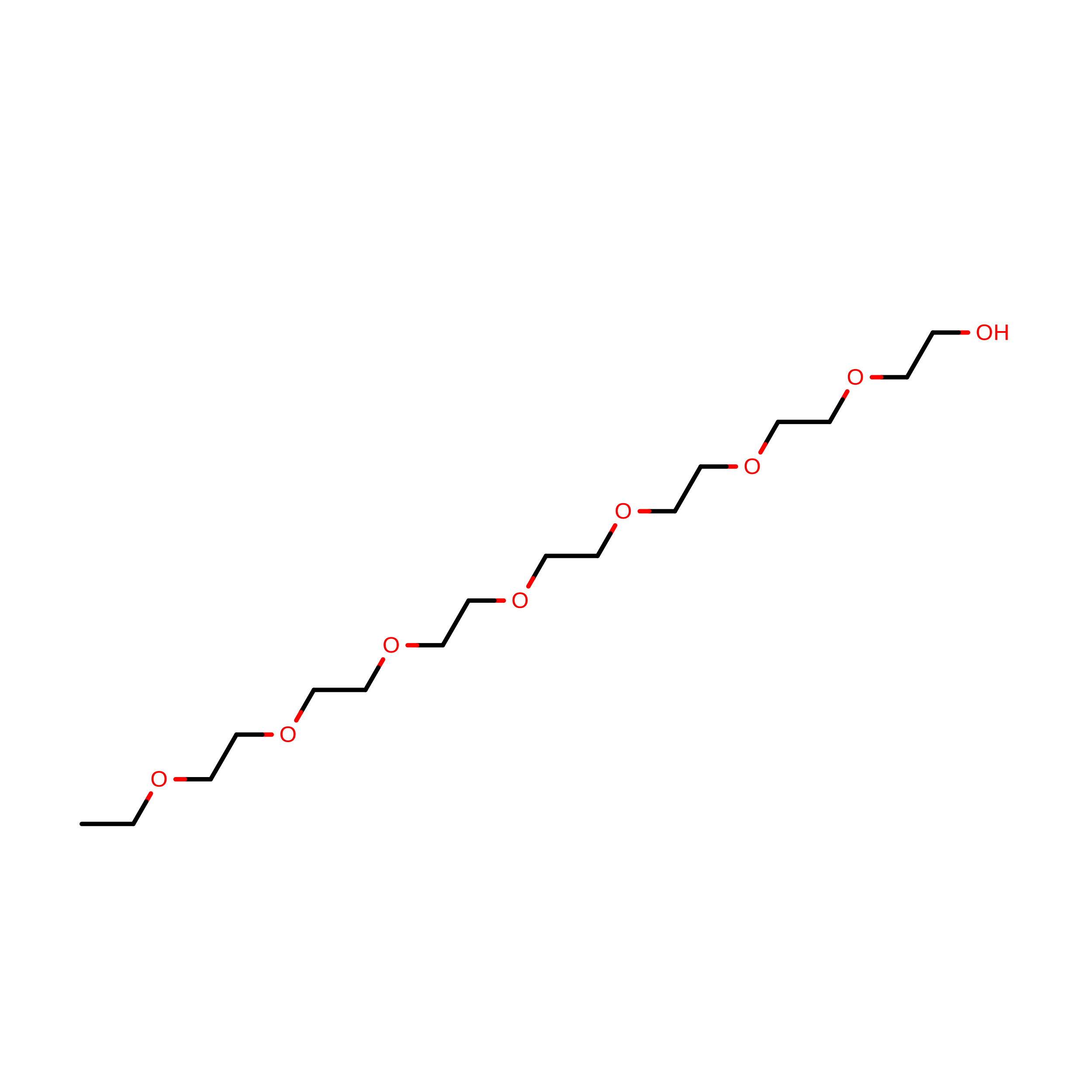
PE4

Query on PE4


Download:Ideal Coordinates CCD File
I [auth B]2-{2-[2-(2-{2-[2-(2-ETHOXY-ETHOXY)-ETHOXY]-ETHOXY}-ETHOXY)-ETHOXY]-ETHOXY}-ETHANOL
C16 H34 O8
PJWQOENWHPEPKI-UHFFFAOYSA-N
PEG

Query on PEG


Download:Ideal Coordinates CCD File
C [auth A]
D [auth A]
E [auth A]
F [auth B]
G [auth B]
C [auth A],
D [auth A],
E [auth A],
F [auth B],
G [auth B],
H [auth B]
DI(HYDROXYETHYL)ETHER
C4 H10 O3
MTHSVFCYNBDYFN-UHFFFAOYSA-N
Modified Residues  2 Unique
IDChains TypeFormula2D DiagramParent
MSE
Query on MSE
A
L-PEPTIDE LINKINGC5 H11 N O2 SeMET
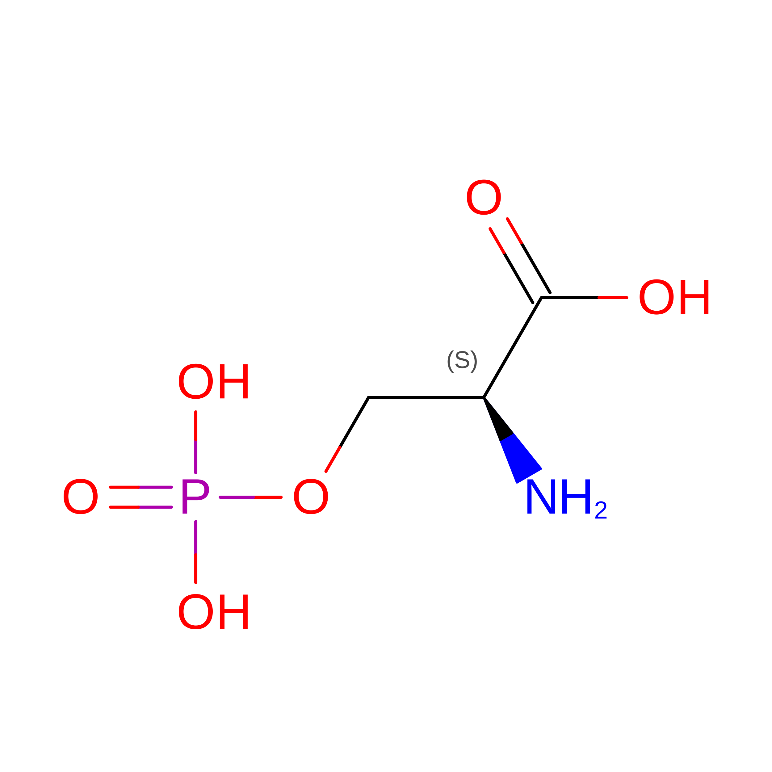
SEP
Query on SEP
B
L-PEPTIDE LINKINGC3 H8 N O6 PSER

Experimental Data & Validation

Experimental Data

  • Method: X-RAY DIFFRACTION
  • Resolution: 1.82 Å
  • R-Value Free:  0.201 (Depositor), 0.203 (DCC) 
  • R-Value Work:  0.176 (Depositor), 0.177 (DCC) 
  • R-Value Observed: 0.177 (Depositor) 
Space Group: P 21 21 21
Unit Cell:
Length ( Å )Angle ( ˚ )
a = 64.991α = 90
b = 69.929β = 90
c = 105.778γ = 90
Software Package:
Software NamePurpose
PHENIXrefinement
PDB_EXTRACTdata extraction
HKL-2000data reduction
HKL-2000data scaling
BALBESphasing

Structure Validation

View Full Validation Report


Entry History 

Revision History  (Full details and data files)

  • Version 1.0: 2013-06-19
    Type: Initial release
  • Version 1.1: 2013-12-25
    Changes: Structure summary
  • Version 1.2: 2023-09-20
    Changes: Data collection, Database references, Derived calculations, Refinement description
  • Version 1.3: 2023-12-06
    Changes: Data collection
  • Version 1.4: 2024-10-16
    Changes: Structure summary