4L1E | pdb_00004l1e

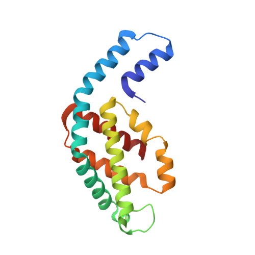
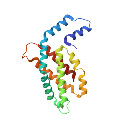
Crystal structure of C-Phycocyanin from Leptolyngbya sp. N62DM


Experimental Data Snapshot

  • Method: X-RAY DIFFRACTION
  • Resolution: 2.61 Å
  • R-Value Free: 
    0.281 (Depositor), 0.282 (DCC) 
  • R-Value Work: 
    0.239 (Depositor), 0.241 (DCC) 
  • R-Value Observed: 
    0.241 (Depositor) 

Starting Model: experimental
View more details

wwPDB Validation 3D Report Full Report

Validation slider image for 4L1E

Ligand Structure Quality Assessment 


This is version 1.3 of the entry. See complete history

Literature

Crystal Structure and Interaction of Phycocyanin with beta-Secretase: A Putative Therapy for Alzheimer's Disease.

Singh, N.K.Hasan, S.S.Kumar, J.Raj, I.Pathan, A.A.Parmar, A.Shakil, S.Gourinath, S.Madamwar, D.

(2014) CNS Neurol Disord Drug Targets 13: 691-698

  • DOI: https://doi.org/10.2174/1871527313666140228114456
  • Primary Citation Related Structures: 
    4L1E

  • PubMed Abstract: 

    Alzheimer's disease (AD) represents a neurological disorder, which is caused by enzymatic degradation of an amyloid precursor protein into short peptide fragments that undergo association to form insoluble plaques. Preliminary studies suggest that cyanobacterial extracts, especially the light-harvesting protein phycocyanin, may provide a means to control the progression of the disease. However, the molecular mechanism of disease control remains elusive. In the present study, intact hexameric phycocyanin was isolated and crystallized from the cyanobacterium Leptolyngbya sp. N62DM, and the structure was solved to a resolution of 2.6 A. Molecular docking studies show that the phycocyanin αβ-dimer interacts with the enzyme β-secretase, which catalyzes the proteolysis of the amyloid precursor protein to form plaques. The molecular docking studies suggest that the interaction between phycocyanin and β-secretase is energetically more favorable than previously reported inhibitor-β-secretase interactions. Transgenic Caenorhabditis elegans worms, with a genotype to serve as an AD-model, were significantly protected by phycocyanin. Therefore, the present study provides a novel structure-based molecular mechanism of phycocyanin-mediated therapy against AD.


  • Organizational Affiliation
    • (Datta Madamwar) BRD School of Biosciences, Sardar Patel Maidan, Vadtal Road, Satellite Campus, Post Box No. 39, Sardar Patel University, Vallabh Vidyanagar-388 120, Gujarat, India. datta_madamwar@yahoo.com.

Macromolecule Content 

  • Total Structure Weight: 224.62 kDa 
  • Atom Count: 15,964 
  • Modeled Residue Count: 2,004 
  • Deposited Residue Count: 2,004 
  • Unique protein chains: 2

Macromolecules

Find similar proteins by:|  3D Structure
Entity ID: 1
MoleculeChains  Sequence LengthOrganismDetailsImage
Phycocyanin alpha chain
A, C, E, G, I
A, C, E, G, I, K
162Leptolyngbya sp. N62DMMutation(s): 0 
UniProt
Find proteins for A0A067XG68 (Leptolyngbya sp. N62DM)
Explore A0A067XG68 
Go to UniProtKB:  A0A067XG68
Entity Groups
Sequence Clusters30% Identity50% Identity70% Identity90% Identity95% Identity100% Identity
UniProt GroupA0A067XG68
Sequence Annotations
Expand
Reference Sequence
Find similar proteins by:|  3D Structure
Entity ID: 2
MoleculeChains  Sequence LengthOrganismDetailsImage
Phycocyanin beta chain
B, D, F, H, J
B, D, F, H, J, L
172Leptolyngbya sp. N62DMMutation(s): 0 
UniProt
Find proteins for A0A067XG69 (Leptolyngbya sp. N62DM)
Explore A0A067XG69 
Go to UniProtKB:  A0A067XG69
Entity Groups
Sequence Clusters30% Identity50% Identity70% Identity90% Identity95% Identity100% Identity
UniProt GroupA0A067XG69
Sequence Annotations
Expand
Reference Sequence

Small Molecules

Ligands 2 Unique
IDChains Name / Formula / InChI Key2D Diagram3D Interactions
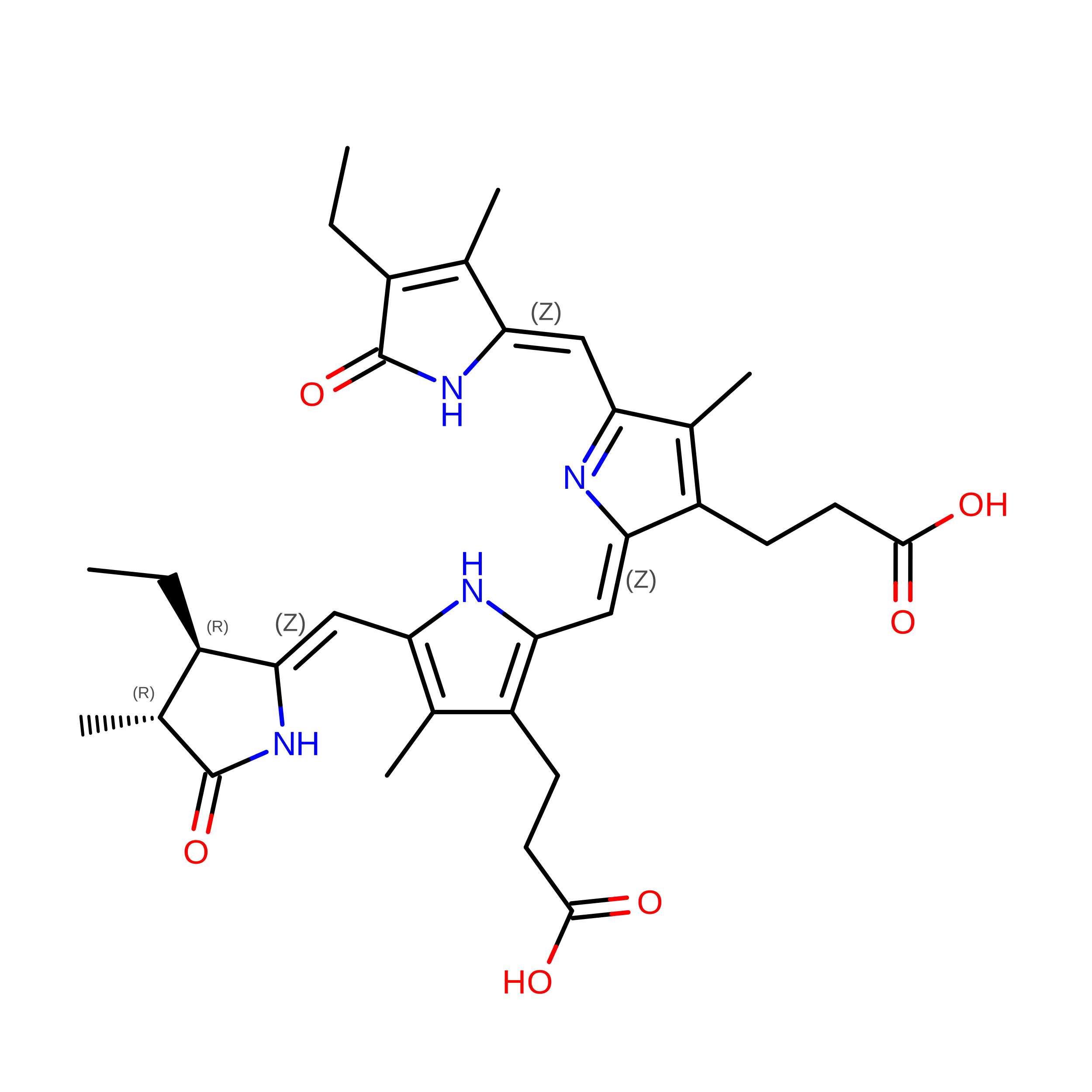
CYC

Query on CYC


Download:Ideal Coordinates CCD File
BA [auth K],
M [auth A],
P [auth C],
S [auth E],
V [auth G]
PHYCOCYANOBILIN
C33 H40 N4 O6
VXTXPYZGDQPMHK-GMXXPEQVSA-N
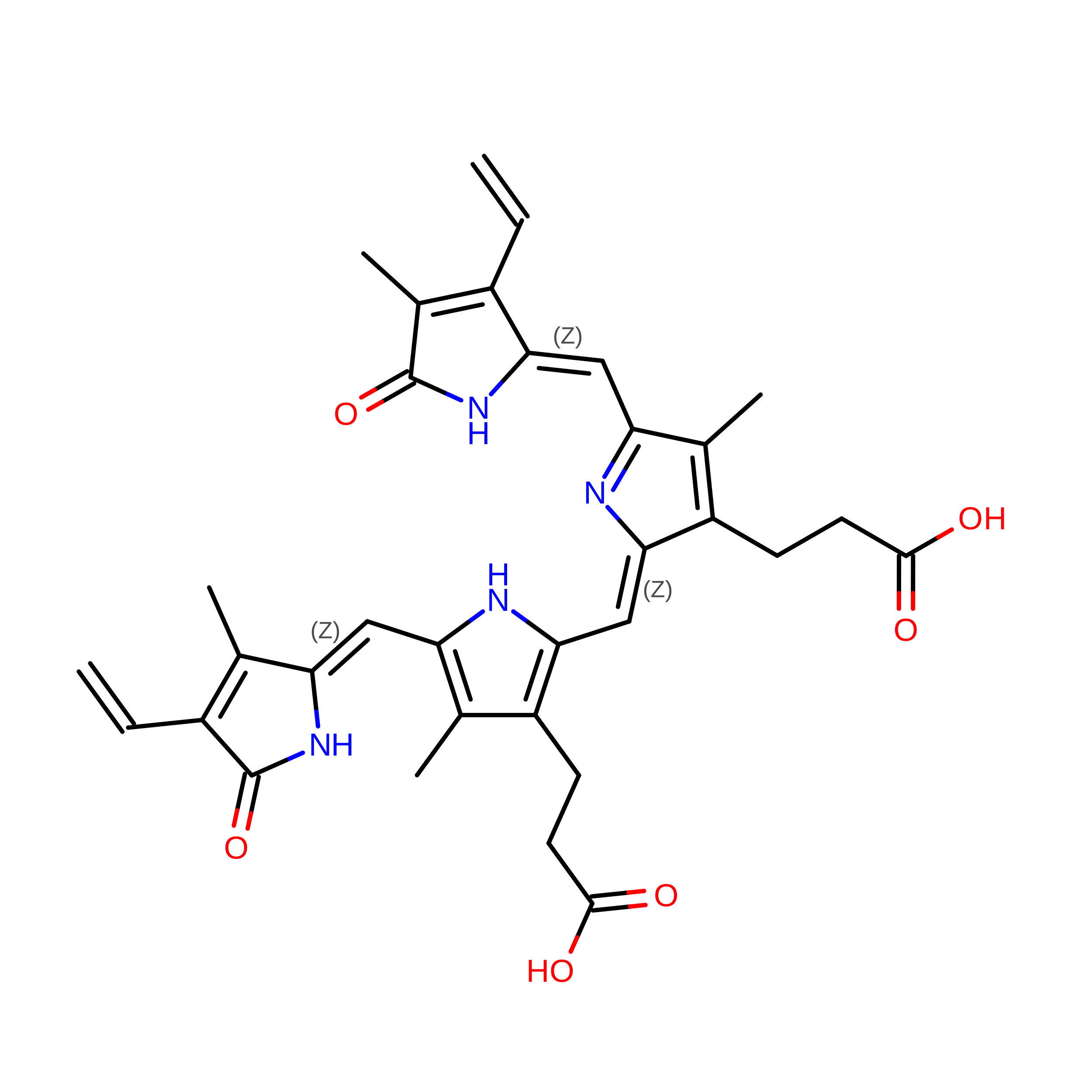
BLA

Query on BLA


Download:Ideal Coordinates CCD File
AA [auth J]
CA [auth L]
DA [auth L]
N [auth B]
O [auth B]
AA [auth J],
CA [auth L],
DA [auth L],
N [auth B],
O [auth B],
Q [auth D],
R [auth D],
T [auth F],
U [auth F],
W [auth H],
X [auth H],
Y [auth I],
Z [auth J]
BILIVERDINE IX ALPHA
C33 H34 N4 O6
GWZYPXHJIZCRAJ-SRVCBVSDSA-N

Experimental Data & Validation

Experimental Data

  • Method: X-RAY DIFFRACTION
  • Resolution: 2.61 Å
  • R-Value Free:  0.281 (Depositor), 0.282 (DCC) 
  • R-Value Work:  0.239 (Depositor), 0.241 (DCC) 
  • R-Value Observed: 0.241 (Depositor) 
Space Group: C 1 2 1
Unit Cell:
Length ( Å )Angle ( ˚ )
a = 183.467α = 90
b = 107.107β = 98.27
c = 111.089γ = 90
Software Package:
Software NamePurpose
PHENIXrefinement
PDB_EXTRACTdata extraction
MAR345dtbdata collection
AUTOMARdata reduction
PHASERphasing

Structure Validation

View Full Validation Report


Ligand Structure Quality Assessment 


Entry History 

Deposition Data

Revision History  (Full details and data files)

  • Version 1.0: 2014-05-21
    Type: Initial release
  • Version 1.1: 2014-05-28
    Changes: Database references
  • Version 1.2: 2014-07-23
    Changes: Database references
  • Version 1.3: 2023-09-20
    Changes: Data collection, Database references, Derived calculations, Refinement description