4L0Y | pdb_00004l0y

Crystal structure of Runx1 and Ets1 bound to TCR alpha promoter (crystal form 1)


Experimental Data Snapshot

  • Method: X-RAY DIFFRACTION
  • Resolution: 2.50 Å
  • R-Value Free: 
    0.245 (Depositor), 0.245 (DCC) 
  • R-Value Work: 
    0.225 (Depositor), 0.224 (DCC) 
  • R-Value Observed: 
    0.225 (Depositor) 

wwPDB Validation 3D Report Full Report

Validation slider image for 4L0Y

This is version 1.3 of the entry. See complete history

Literature

Structural basis of Ets1 activation by Runx1.

Shrivastava, T.Mino, K.Babayeva, N.D.Baranovskaya, O.I.Rizzino, A.Tahirov, T.H.

(2014) Leukemia 28: 2040-2048

  • DOI: https://doi.org/10.1038/leu.2014.111
  • Primary Citation Related Structures: 
    4L0Y, 4L0Z, 4L18

  • PubMed Abstract: 

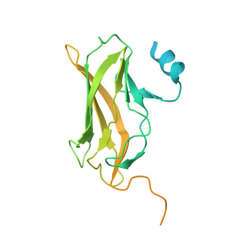
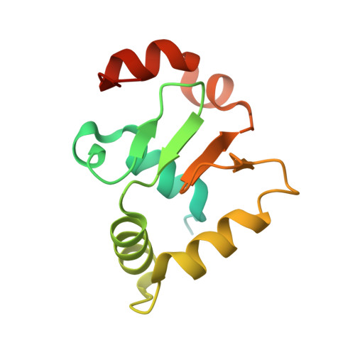
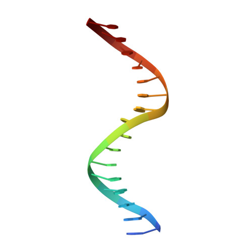
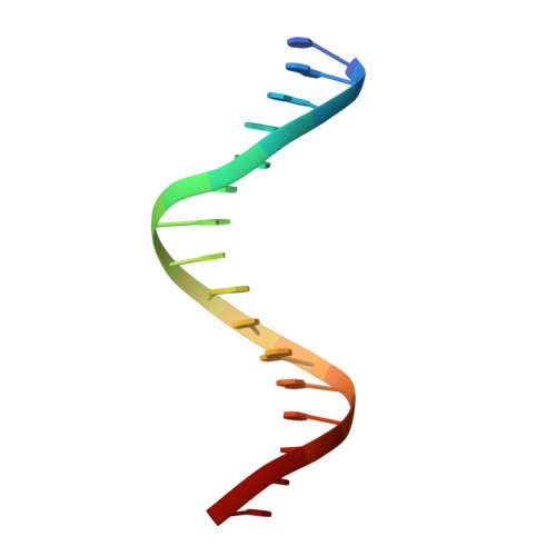
    Runx1 is required for definitive hematopoiesis and is well known for its frequent chromosomal translocations and point mutations in leukemia. Runx1 regulates a variety of genes via Ets1 activation on an Ets1•Runx1 composite DNA sequence. The structural basis of such regulation remains unresolved. To address this problem, we determined the crystal structure of the ternary complex containing Runx1(1-242) and Ets1(296-441) bound to T-cell receptor alpha (TCRα) enhancer DNA. In the crystal, an Ets1-interacting domain of Runx1 is bound to the Ets1 DNA-binding domain and displaced an entire autoinhibitory module of Ets1, revealing a novel mechanism of Ets1 activation. The DNA-binding and transcriptional studies with a variety of structure-guided Runx1 mutants confirmed a critical role of direct Ets1•Runx1 interaction in Ets1 activation. More importantly, the discovered mechanism provides a plausible explanation for how the Ets1•Runx1 interaction effectively activates not only a wild-type Ets1, but also a highly inhibited phosphorylated form of Ets1.


  • Organizational Affiliation
    • Eppley Institute for Research in Cancer and Allied Diseases, University of Nebraska Medical Center, Omaha, NE, USA.

Macromolecule Content 

  • Total Structure Weight: 53.26 kDa 
  • Atom Count: 2,584 
  • Modeled Residue Count: 264 
  • Deposited Residue Count: 420 
  • Unique protein chains: 2
  • Unique nucleic acid chains: 2

Macromolecules


Find similar proteins by:|  3D Structure
Entity ID: 1
MoleculeChains  Sequence LengthOrganismDetailsImage
Runt-related transcription factor 1242Mus musculusMutation(s): 0 
Gene Names: Runx1Aml1Cbfa2Pebp2ab
UniProt
Find proteins for Q03347 (Mus musculus)
Explore Q03347 
Go to UniProtKB:  Q03347
Entity Groups
Sequence Clusters30% Identity50% Identity70% Identity90% Identity95% Identity100% Identity
UniProt GroupQ03347
Sequence Annotations
Expand
Reference Sequence
Find similar proteins by:|  3D Structure
Entity ID: 2
MoleculeChains  Sequence LengthOrganismDetailsImage
Protein C-ets-1146Homo sapiensMutation(s): 0 
Gene Names: ETS1EWSR2
UniProt & NIH Common Fund Data Resources
Find proteins for P14921 (Homo sapiens)
Explore P14921 
Go to UniProtKB:  P14921
PHAROS:  P14921
GTEx:  ENSG00000134954 
Entity Groups
Sequence Clusters30% Identity50% Identity70% Identity90% Identity95% Identity100% Identity
UniProt GroupP14921
Sequence Annotations
Expand
Reference Sequence
Find similar nucleic acids by:  Sequence
Entity ID: 3
MoleculeChains LengthOrganismImage
5'-D(*GP*GP*AP*AP*GP*CP*CP*AP*CP*AP*TP*CP*CP*TP*CP*T)-3'16Homo sapiens
Sequence Annotations
Expand
Reference Sequence
Find similar nucleic acids by:  Sequence
Entity ID: 4
MoleculeChains LengthOrganismImage
5'-D(*CP*AP*GP*AP*GP*GP*AP*TP*GP*TP*GP*GP*CP*TP*TP*C)-3'16Homo sapiens
Sequence Annotations
Expand
Reference Sequence

Experimental Data & Validation

Experimental Data

  • Method: X-RAY DIFFRACTION
  • Resolution: 2.50 Å
  • R-Value Free:  0.245 (Depositor), 0.245 (DCC) 
  • R-Value Work:  0.225 (Depositor), 0.224 (DCC) 
  • R-Value Observed: 0.225 (Depositor) 
Space Group: C 2 2 21
Unit Cell:
Length ( Å )Angle ( ˚ )
a = 102.622α = 90
b = 138.945β = 90
c = 98.439γ = 90
Software Package:
Software NamePurpose
ADSCdata collection
CNSrefinement
HKL-2000data reduction
HKL-2000data scaling
CNSphasing

Structure Validation

View Full Validation Report


Entry History 

Deposition Data

Revision History  (Full details and data files)

  • Version 1.0: 2014-03-26
    Type: Initial release
  • Version 1.1: 2014-04-02
    Changes: Database references
  • Version 1.2: 2014-10-22
    Changes: Database references
  • Version 1.3: 2024-02-28
    Changes: Data collection, Database references