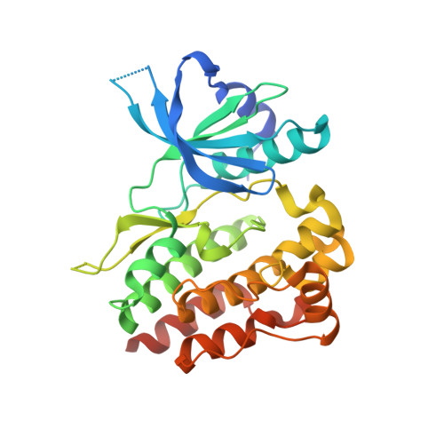
4L01 | pdb_00004l01

Crystal structure of the V658F apo Jak1 pseudokinase domain


Experimental Data Snapshot

  • Method: X-RAY DIFFRACTION
  • Resolution: 1.90 Å
  • R-Value Free: 
    0.233 (Depositor), 0.233 (DCC) 
  • R-Value Work: 
    0.202 (Depositor), 0.202 (DCC) 
  • R-Value Observed: 
    0.204 (Depositor) 

wwPDB Validation 3D Report Full Report

Validation slider image for 4L01

This is version 1.3 of the entry. See complete history. 

Literature

Structure of a pseudokinase-domain switch that controls oncogenic activation of Jak kinases.

Toms, A.V., Deshpande, A., McNally, R., Jeong, Y., Rogers, J.M., Kim, C.U., Gruner, S.M., Ficarro, S.B., Marto, J.A., Sattler, M., Griffin, J.D., Eck, M.J.

(2013) Nat Struct Mol Biol 20: 1221-1223

  • DOI: https://doi.org/10.1038/nsmb.2673
  • Primary Citation Related Structures: 
    4L00, 4L01

  • PubMed Abstract: 

    The V617F mutation in the Jak2 pseudokinase domain causes myeloproliferative neoplasms, and the equivalent mutation in Jak1 (V658F) is found in T-cell leukemias. Crystal structures of wild-type and V658F-mutant human Jak1 pseudokinase reveal a conformational switch that remodels a linker segment encoded by exon 12, which is also a site of mutations in Jak2. This switch is required for V617F-mediated Jak2 activation and possibly for physiologic Jak activation.


  • Organizational Affiliation: 
    • 1] Department of Biological Chemistry and Molecular Pharmacology, Harvard Medical School, Boston, Massachusetts, USA. [2] Department of Cancer Biology, Dana-Farber Cancer Institute, Boston, Massachusetts, USA.

Macromolecule Content 

  • Total Structure Weight: 69.93 kDa 
  • Atom Count: 4,700 
  • Modeled Residue Count: 548 
  • Deposited Residue Count: 608 
  • Unique protein chains: 1

Macromolecules

Find similar proteins by:|  3D Structure
Entity ID: 1
MoleculeChains  Sequence LengthOrganismDetailsImage
Tyrosine-protein kinase JAK1
A, B
304Homo sapiensMutation(s): 1 
Gene Names: JAK1, JAK1A, JAK1B
EC: 2.7.10.2
UniProt & NIH Common Fund Data Resources
Find proteins for P23458 (Homo sapiens)
Explore P23458 
Go to UniProtKB:  P23458
PHAROS:  P23458
GTEx:  ENSG00000162434 
Entity Groups
Sequence Clusters30% Identity50% Identity70% Identity90% Identity95% Identity100% Identity
UniProt GroupP23458
Sequence Annotations
Expand
Reference Sequence

Experimental Data & Validation

Experimental Data

  • Method: X-RAY DIFFRACTION
  • Resolution: 1.90 Å
  • R-Value Free:  0.233 (Depositor), 0.233 (DCC) 
  • R-Value Work:  0.202 (Depositor), 0.202 (DCC) 
  • R-Value Observed: 0.204 (Depositor) 
Space Group: P 1 21 1
Unit Cell:
Length ( Å )Angle ( ˚ )
a = 47.288α = 90
b = 80.557β = 95.29
c = 76.555γ = 90
Software Package:
Software NamePurpose
ADSCdata collection
PHASERphasing
REFMACrefinement
HKL-2000data reduction
HKL-2000data scaling

Structure Validation

View Full Validation Report


Entry History 

Deposition Data

Revision History  (Full details and data files)

  • Version 1.0: 2013-09-04
    Type: Initial release
  • Version 1.1: 2013-10-02
    Changes: Database references
  • Version 1.2: 2014-02-05
    Changes: Database references
  • Version 1.3: 2024-02-28
    Changes: Data collection, Database references