4KZS | pdb_00004kzs

Crystal structure of the secreted protein HP1454 from the human pathogen Helicobacter pylori


Experimental Data Snapshot

  • Method: X-RAY DIFFRACTION
  • Resolution: 2.70 Å
  • R-Value Free: 
    0.304 (Depositor), 0.317 (DCC) 
  • R-Value Work: 
    0.248 (Depositor), 0.260 (DCC) 
  • R-Value Observed: 
    0.250 (Depositor) 

wwPDB Validation 3D Report Full Report

Validation slider image for 4KZS

This is version 1.3 of the entry. See complete history

Literature

Crystal structure of the secreted protein HP1454 from the human pathogen Helicobacter pylori.

Quarantini, S.Cendron, L.Zanotti, G.

(2014) Proteins 82: 2868-2873

  • DOI: https://doi.org/10.1002/prot.24608
  • Primary Citation Related Structures: 
    4KZS

  • PubMed Abstract: 

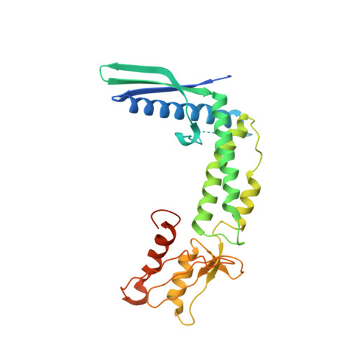
    HP1454 is a protein of 303 amino acids found in the extracellular milieu of Helicobacter pylori. The protein structure, crystallized in the orthorhombic C222₁ space group with one molecule per asymmetric unit, has been determined using the single-wavelength anomalous dispersion method. HP1454 exhibits an elongated bent shape, composed of three distinct domains. Each domain possesses a fold already present in other structures: Domain I contains a three-strand antiparallel β-barrel flanked by a long α-helix, Domain II is an anti-parallel three-helix bundle, and Domain III a β-sheet flanked by two α-helices. The overall assembly of the protein does not bear any similarity with known structures.


  • Organizational Affiliation
    • Department of Biomedical Sciences, University of Padua, Viale G. Colombo 3, Padua, 35131, Italy.

Macromolecule Content 

  • Total Structure Weight: 32.26 kDa 
  • Atom Count: 1,961 
  • Modeled Residue Count: 242 
  • Deposited Residue Count: 285 
  • Unique protein chains: 1

Macromolecules

Find similar proteins by:|  3D Structure
Entity ID: 1
MoleculeChains  Sequence LengthOrganismDetailsImage
LPP20 lipofamily protein285Helicobacter pylori R046WaMutation(s): 0 
Gene Names: hp1454OUO_1490

Small Molecules

Ligands 1 Unique
IDChains Name / Formula / InChI Key2D Diagram3D Interactions
1PE

Query on 1PE


Download:Ideal Coordinates CCD File
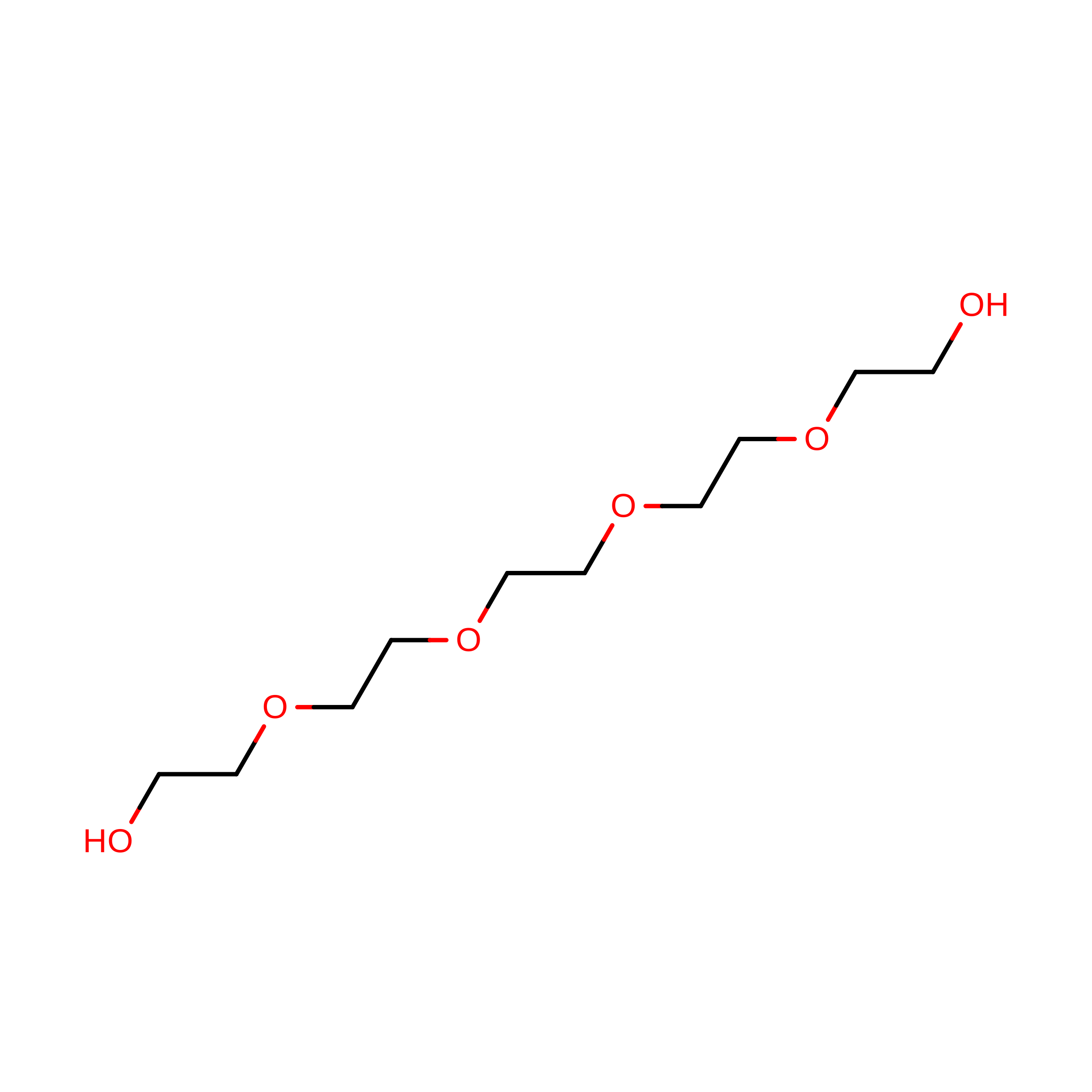
B [auth A]PENTAETHYLENE GLYCOL
C10 H22 O6
JLFNLZLINWHATN-UHFFFAOYSA-N

Experimental Data & Validation

Experimental Data

  • Method: X-RAY DIFFRACTION
  • Resolution: 2.70 Å
  • R-Value Free:  0.304 (Depositor), 0.317 (DCC) 
  • R-Value Work:  0.248 (Depositor), 0.260 (DCC) 
  • R-Value Observed: 0.250 (Depositor) 
Space Group: C 2 2 21
Unit Cell:
Length ( Å )Angle ( ˚ )
a = 73.64α = 90
b = 101.35β = 90
c = 121.08γ = 90
Software Package:
Software NamePurpose
MAR345data collection
SHARPphasing
REFMACrefinement
XDSdata reduction
SCALAdata scaling

Structure Validation

View Full Validation Report


Entry History 

Deposition Data

Revision History  (Full details and data files)

  • Version 1.0: 2014-06-04
    Type: Initial release
  • Version 1.1: 2014-10-22
    Changes: Database references
  • Version 1.2: 2017-11-15
    Changes: Refinement description
  • Version 1.3: 2024-02-28
    Changes: Data collection, Database references, Derived calculations