4KZC | pdb_00004kzc

Structure of PI3K gamma with Imidazopyridine inhibitors


Experimental Data Snapshot

  • Method: X-RAY DIFFRACTION
  • Resolution: 3.25 Å
  • R-Value Free: 
    0.256 (Depositor), 0.270 (DCC) 
  • R-Value Work: 
    0.235 (Depositor), 0.255 (DCC) 
  • R-Value Observed: 
    0.236 (Depositor) 

wwPDB Validation 3D Report Full Report

Validation slider image for 4KZC

Ligand Structure Quality Assessment 


This is version 1.2 of the entry. See complete history

Literature

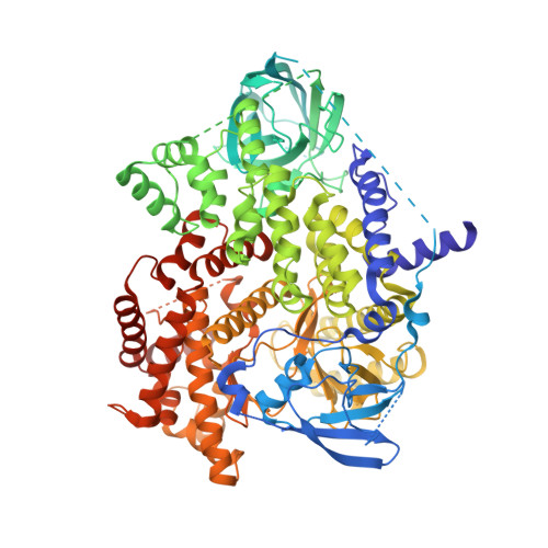
Structure guided optimization of a fragment hit to imidazopyridine inhibitors of PI3K.

Pecchi, S.Ni, Z.J.Han, W.Smith, A.Lan, J.Burger, M.Merritt, H.Wiesmann, M.Chan, J.Kaufman, S.Knapp, M.S.Janssen, J.Huh, K.Voliva, C.F.

(2013) Bioorg Med Chem Lett 23: 4652-4656

  • DOI: https://doi.org/10.1016/j.bmcl.2013.06.010
  • Primary Citation Related Structures: 
    4KZ0, 4KZC

  • PubMed Abstract: 

    PI3 kinases are a family of lipid kinases mediating numerous cell processes such as proliferation, migration and differentiation. The PI3 Kinase pathway is often de-regulated in cancer through PI3Kα overexpression, gene amplification, mutations and PTEN phosphatase deletion. PI3K inhibitors represent therefore an attractive therapeutic modality for cancer treatment. Herein we describe how the potency of a benzothiazole fragment hit was quickly improved based on structural information and how this early chemotype was further optimized through scaffold hopping. This effort led to the identification of a series of 2-acetamido-5-heteroaryl imidazopyridines showing potent in vitro activity against all class I PI3Ks and attractive pharmacokinetic properties.


  • Organizational Affiliation
    • Global Discovery Chemistry/Oncology and Exploratory Chemistry, Novartis Institutes for Biomedical Research, Emeryville, CA, USA.

Macromolecule Content 

  • Total Structure Weight: 111.19 kDa 
  • Atom Count: 6,282 
  • Modeled Residue Count: 821 
  • Deposited Residue Count: 966 
  • Unique protein chains: 1

Macromolecules

Find similar proteins by:|  3D Structure
Entity ID: 1
MoleculeChains  Sequence LengthOrganismDetailsImage
Phosphatidylinositol 4,5-bisphosphate 3-kinase catalytic subunit gamma isoform966Homo sapiensMutation(s): 1 
Gene Names: PIK3CG
EC: 2.7.1.153 (PDB Primary Data), 2.7.11.1 (PDB Primary Data), 2.7.1.137 (UniProt), 2.7.1.154 (UniProt)
UniProt & NIH Common Fund Data Resources
Find proteins for P48736 (Homo sapiens)
Explore P48736 
Go to UniProtKB:  P48736
PHAROS:  P48736
GTEx:  ENSG00000105851 
Entity Groups
Sequence Clusters30% Identity50% Identity70% Identity90% Identity95% Identity100% Identity
UniProt GroupP48736
Sequence Annotations
Expand
Reference Sequence

Small Molecules

Ligands 2 Unique
IDChains Name / Formula / InChI Key2D Diagram3D Interactions
1UK

Query on 1UK


Download:Ideal Coordinates CCD File
B [auth A]N-{6-[6-amino-5-(trifluoromethyl)pyridin-3-yl]imidazo[1,2-a]pyridin-2-yl}acetamide
C15 H12 F3 N5 O
TVUPSIBSWDXJGQ-UHFFFAOYSA-N
SO4

Query on SO4


Download:Ideal Coordinates CCD File
C [auth A]SULFATE ION
O4 S
QAOWNCQODCNURD-UHFFFAOYSA-L

Experimental Data & Validation

Experimental Data

  • Method: X-RAY DIFFRACTION
  • Resolution: 3.25 Å
  • R-Value Free:  0.256 (Depositor), 0.270 (DCC) 
  • R-Value Work:  0.235 (Depositor), 0.255 (DCC) 
  • R-Value Observed: 0.236 (Depositor) 
Space Group: C 1 2 1
Unit Cell:
Length ( Å )Angle ( ˚ )
a = 146.383α = 90
b = 68.313β = 95.24
c = 108.161γ = 90
Software Package:
Software NamePurpose
SCALAdata scaling
TNTrefinement
PDB_EXTRACTdata extraction
BOSdata collection
XSCALEdata scaling
PHASERphasing
BUSTERrefinement

Structure Validation

View Full Validation Report


Ligand Structure Quality Assessment 


Entry History 

Deposition Data

Revision History  (Full details and data files)

  • Version 1.0: 2013-07-17
    Type: Initial release
  • Version 1.1: 2013-08-28
    Changes: Database references
  • Version 1.2: 2024-02-28
    Changes: Data collection, Database references, Derived calculations