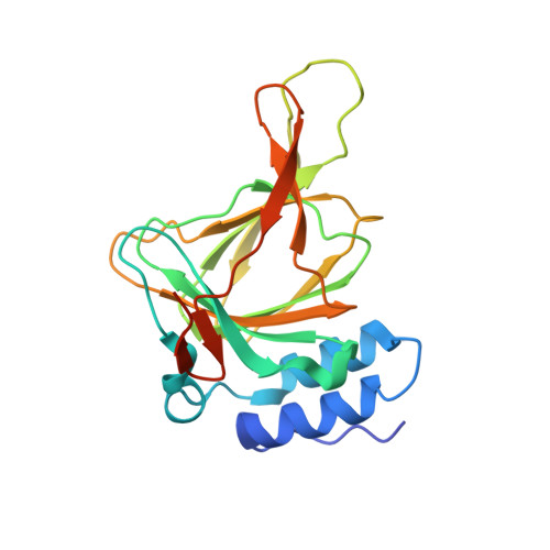
Mechanistic implications of persulfenate and persulfide binding in the active site of cysteine dioxygenase.
Souness, R.J., Kleffmann, T., Tchesnokov, E.P., Wilbanks, S.M., Jameson, G.B., Jameson, G.N.(2013) Biochemistry 52: 7606-7617
- PubMed: 24084026 Search on PubMed
- DOI: https://doi.org/10.1021/bi400661a
- Primary Citation Related Structures:
4KWJ, 4KWK, 4KWL - PubMed Abstract:
Describing the organization of substrates and substrate analogues in the active site of cysteine dioxygenase identifies potential intermediates in this critical yet poorly understood reaction, the oxidation of cysteine to cysteine sulfinic acid. The fortuitous formation of persulfides under crystallization conditions has allowed their binding in the active site of cysteine dioxygenase to be studied. The crystal structures of cysteine persulfide and 3-mercaptopropionic acid persulfide bound to iron(II) in the active site show that binding of the persulfide occurs via the distal sulfide and, in the case of the cysteine persulfide, the amine also binds. Persulfide was detected by mass spectrometry in both the crystal and the drop, suggesting its origin is chemical rather than enzymatic. A mechanism involving the formation of the relevant disulfide from sulfide produced by hydrolysis of dithionite is proposed. In comparison, persulfenate {observed bound to cysteine dioxygenase [Simmons, C. R., et al. (2008) Biochemistry 47, 11390]} is shown through mass spectrometry to occur only in the crystal and not in the surrounding drop, suggesting that in the crystalline state the persulfenate does not lie on the reaction pathway. Stabilization of both the persulfenate and the persulfides does, however, suggest the position in which dioxygen binds during catalysis.
- Department of Chemistry and MacDiarmid Institute for Advanced Materials and Nanotechnology and ‡Department of Biochemistry, University of Otago , P.O. Box 56, Dunedin 9054, New Zealand.
Organizational Affiliation: