4KRR | pdb_00004krr

Crystal structure of Drosophila WntD N-terminal domain-linker (residues 31-240)


Experimental Data Snapshot

  • Method: X-RAY DIFFRACTION
  • Resolution: 2.12 Å
  • R-Value Free: 
    0.203 (Depositor), 0.206 (DCC) 
  • R-Value Work: 
    0.179 (Depositor), 0.184 (DCC) 
  • R-Value Observed: 
    0.180 (Depositor) 

Starting Model: experimental
View more details

wwPDB Validation 3D Report Full Report

Validation slider image for 4KRR

This is version 1.4 of the entry. See complete history

Literature

Structural Studies of Wnts and Identification of an LRP6 Binding Site.

Chu, M.L.Ahn, V.E.Choi, H.J.Daniels, D.L.Nusse, R.Weis, W.I.

(2013) Structure 21: 1235-1242

  • DOI: https://doi.org/10.1016/j.str.2013.05.006
  • Primary Citation Related Structures: 
    4KRR

  • PubMed Abstract: 

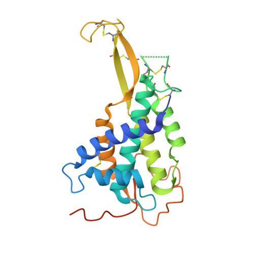
    Wnts are secreted growth factors that have critical roles in cell fate determination and stem cell renewal. The Wnt/β-catenin pathway is initiated by binding of a Wnt protein to a Frizzled (Fzd) receptor and a coreceptor, LDL receptor-related protein 5 or 6 (LRP5/6). We report the 2.1 Å resolution crystal structure of a Drosophila WntD fragment encompassing the N-terminal domain and the linker that connects it to the C-terminal domain. Differences in the structures of WntD and Xenopus Wnt8, including the positions of a receptor-binding β hairpin and a large solvent-filled cavity in the helical core, indicate conformational plasticity in the N-terminal domain that may be important for Wnt-Frizzled specificity. Structure-based mutational analysis of mouse Wnt3a shows that the linker between the N- and C-terminal domains is required for LRP6 binding. These findings provide important insights into Wnt function and evolution.


  • Organizational Affiliation
    • Departments of Structural Biology and Molecular and Cellular Physiology, Stanford University School of Medicine, Stanford, CA 94305, USA.

Macromolecule Content 

  • Total Structure Weight: 24.92 kDa 
  • Atom Count: 1,562 
  • Modeled Residue Count: 194 
  • Deposited Residue Count: 221 
  • Unique protein chains: 1

Macromolecules

Find similar proteins by:|  3D Structure
Entity ID: 1
MoleculeChains  Sequence LengthOrganismDetailsImage
Wnt inhibitor of Dorsal protein221Drosophila melanogasterMutation(s): 0 
Gene Names: CG8458Wnt8wntD
UniProt
Find proteins for Q9VFX1 (Drosophila melanogaster)
Explore Q9VFX1 
Go to UniProtKB:  Q9VFX1
Entity Groups
Sequence Clusters30% Identity50% Identity70% Identity90% Identity95% Identity100% Identity
UniProt GroupQ9VFX1
Sequence Annotations
Expand
Reference Sequence

Experimental Data & Validation

Experimental Data

  • Method: X-RAY DIFFRACTION
  • Resolution: 2.12 Å
  • R-Value Free:  0.203 (Depositor), 0.206 (DCC) 
  • R-Value Work:  0.179 (Depositor), 0.184 (DCC) 
  • R-Value Observed: 0.180 (Depositor) 
Space Group: P 41
Unit Cell:
Length ( Å )Angle ( ˚ )
a = 59.625α = 90
b = 59.625β = 90
c = 66.955γ = 90
Software Package:
Software NamePurpose
PHENIXrefinement
PHASERphasing
CNSrefinement
SCALAdata scaling
PDB_EXTRACTdata extraction
Blu-Icedata collection
XDSdata reduction

Structure Validation

View Full Validation Report


Entry History 

Deposition Data

Revision History  (Full details and data files)

  • Version 1.0: 2013-07-10
    Type: Initial release
  • Version 1.1: 2013-07-17
    Changes: Database references
  • Version 1.2: 2019-07-17
    Changes: Data collection, Refinement description
  • Version 1.3: 2023-09-20
    Changes: Data collection, Database references, Derived calculations, Refinement description
  • Version 1.4: 2024-10-30
    Changes: Structure summary