4KQO | pdb_00004kqo

Crystal structure of penicillin-binding protein 3 from pseudomonas aeruginosa in complex with piperacillin


Experimental Data Snapshot

  • Method: X-RAY DIFFRACTION
  • Resolution: 2.31 Å
  • R-Value Free: 
    0.250 (Depositor), 0.255 (DCC) 
  • R-Value Work: 
    0.217 (Depositor), 0.220 (DCC) 
  • R-Value Observed: 
    0.219 (Depositor) 

Starting Model: experimental
View more details

wwPDB Validation 3D Report Full Report

Validation slider image for 4KQO

Ligand Structure Quality Assessment 


This is version 1.2 of the entry. See complete history

Literature

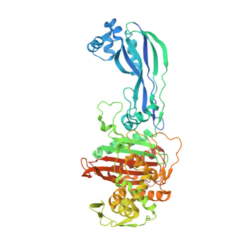
Binding of (5S)-Penicilloic Acid to Penicillin Binding Protein 3.

van Berkel, S.S.Nettleship, J.E.Leung, I.K.Brem, J.Choi, H.Stuart, D.I.Claridge, T.D.McDonough, M.A.Owens, R.J.Ren, J.Schofield, C.J.

(2013) ACS Chem Biol 8: 2112-2116

  • DOI: https://doi.org/10.1021/cb400200h
  • Primary Citation Related Structures: 
    4KQO, 4KQQ, 4KQR

  • PubMed Abstract: 

    β-Lactam antibiotics react with penicillin binding proteins (PBPs) to form relatively stable acyl-enzyme complexes. We describe structures derived from the reaction of piperacillin with PBP3 (Pseudomonas aeruginosa) including not only the anticipated acyl-enzyme complex but also an unprecedented complex with (5S)-penicilloic acid, which was formed by C-5 epimerization of the nascent (5R)-penicilloic acid product. Formation of the complex was confirmed by solution studies, including NMR. Together, these results will be useful in the design of new PBP inhibitors and raise the possibility that noncovalent PBP inhibition by penicilloic acids may be of clinical relevance.


  • Organizational Affiliation
    • Chemistry Research Laboratory, University of Oxford , 12 Mansfield Road, Oxford OX1 3TA, U.K.

Macromolecule Content 

  • Total Structure Weight: 123.76 kDa 
  • Atom Count: 8,008 
  • Modeled Residue Count: 1,003 
  • Deposited Residue Count: 1,128 
  • Unique protein chains: 1

Macromolecules

Find similar proteins by:|  3D Structure
Entity ID: 1
MoleculeChains  Sequence LengthOrganismDetailsImage
Penicillin-binding protein 3
A, B
564Pseudomonas aeruginosa PAO1Mutation(s): 0 
Gene Names: ftsIPA4418
EC: 3.4.16.4
UniProt
Find proteins for G3XD46 (Pseudomonas aeruginosa (strain ATCC 15692 / DSM 22644 / CIP 104116 / JCM 14847 / LMG 12228 / 1C / PRS 101 / PAO1))
Explore G3XD46 
Go to UniProtKB:  G3XD46
Entity Groups
Sequence Clusters30% Identity50% Identity70% Identity90% Identity95% Identity100% Identity
UniProt GroupG3XD46
Sequence Annotations
Expand
Reference Sequence

Small Molecules

Ligands 4 Unique
IDChains Name / Formula / InChI Key2D Diagram3D Interactions
JPP

Query on JPP


Download:Ideal Coordinates CCD File
C [auth A],
F [auth B]
Piperacillin (Open Form)
C23 H29 N5 O7 S
CJYMHKMECTVNSA-XLMAVXFVSA-N
GOL

Query on GOL


Download:Ideal Coordinates CCD File
D [auth A],
E [auth A],
G [auth B]
GLYCEROL
C3 H8 O3
PEDCQBHIVMGVHV-UHFFFAOYSA-N
IMD

Query on IMD


Download:Ideal Coordinates CCD File
H [auth B]IMIDAZOLE
C3 H5 N2
RAXXELZNTBOGNW-UHFFFAOYSA-O
CL

Query on CL


Download:Ideal Coordinates CCD File
I [auth B],
J [auth B]
CHLORIDE ION
Cl
VEXZGXHMUGYJMC-UHFFFAOYSA-M

Experimental Data & Validation

Experimental Data

  • Method: X-RAY DIFFRACTION
  • Resolution: 2.31 Å
  • R-Value Free:  0.250 (Depositor), 0.255 (DCC) 
  • R-Value Work:  0.217 (Depositor), 0.220 (DCC) 
  • R-Value Observed: 0.219 (Depositor) 
Space Group: P 1
Unit Cell:
Length ( Å )Angle ( ˚ )
a = 57.049α = 71.26
b = 74.935β = 86.04
c = 82.785γ = 85.59
Software Package:
Software NamePurpose
MOLREPphasing
REFMACrefinement
HKL-2000data reduction
SCALEPACKdata scaling

Structure Validation

View Full Validation Report


Ligand Structure Quality Assessment 


Entry History 

Deposition Data

Revision History  (Full details and data files)

  • Version 1.0: 2013-11-06
    Type: Initial release
  • Version 1.1: 2023-09-20
    Changes: Data collection, Database references, Derived calculations, Refinement description
  • Version 1.2: 2024-10-30
    Changes: Structure summary