4KQB | pdb_00004kqb

crystal structure of the Golgi casein kinase with Mn/ADP bound


Experimental Data Snapshot

  • Method: X-RAY DIFFRACTION
  • Resolution: 3.04 Å
  • R-Value Free: 
    0.253 (Depositor), 0.257 (DCC) 
  • R-Value Work: 
    0.215 (Depositor) 
  • R-Value Observed: 
    0.217 (Depositor) 

wwPDB Validation 3D Report Full Report

Validation slider image for 4KQB

Ligand Structure Quality Assessment 


This is version 2.1 of the entry. See complete history

Literature

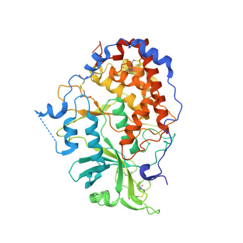
Crystal structure of the Golgi casein kinase.

Xiao, J.Tagliabracci, V.S.Wen, J.Kim, S.A.Dixon, J.E.

(2013) Proc Natl Acad Sci U S A 110: 10574-10579

  • DOI: https://doi.org/10.1073/pnas.1309211110
  • Primary Citation Related Structures: 
    4KQA, 4KQB

  • PubMed Abstract: 

    The family with sequence similarity 20 (Fam20) kinases phosphorylate extracellular substrates and play important roles in biomineralization. Fam20C is the Golgi casein kinase that phosphorylates secretory pathway proteins within Ser-x-Glu/pSer motifs. Mutations in Fam20C cause Raine syndrome, an osteosclerotic bone dysplasia. Here we report the crystal structure of the Fam20C ortholog from Caenorhabditis elegans. The nucleotide-free and Mn/ADP-bound structures unveil an atypical protein kinase-like fold and highlight residues critical for activity. The position of the regulatory αC helix and the lack of an activation loop indicate an architecture primed for efficient catalysis. Furthermore, several distinct elements, including the presence of disulfide bonds, suggest that the Fam20 family diverged early in the evolution of the protein kinase superfamily. Our results reinforce the structural diversity of protein kinases and have important implications for patients with disorders of biomineralization.


  • Organizational Affiliation
    • Department of Pharmacology, University of California at San Diego, La Jolla, CA 92093, USA.

Macromolecule Content 

  • Total Structure Weight: 109.29 kDa 
  • Atom Count: 7,100 
  • Modeled Residue Count: 837 
  • Deposited Residue Count: 906 
  • Unique protein chains: 1

Macromolecules

Find similar proteins by:|  3D Structure
Entity ID: 1
MoleculeChains  Sequence LengthOrganismDetailsImage
Protein H03A11.1
A, B
453Caenorhabditis elegansMutation(s): 0 
Gene Names: CELE_H03A11.1H03A11.1
EC: 2.7.11.1
UniProt
Find proteins for Q9XTW2 (Caenorhabditis elegans)
Explore Q9XTW2 
Go to UniProtKB:  Q9XTW2
Entity Groups
Sequence Clusters30% Identity50% Identity70% Identity90% Identity95% Identity100% Identity
UniProt GroupQ9XTW2
Glycosylation
Glycosylation Sites: 2
Sequence Annotations
Expand
Reference Sequence

Oligosaccharides

Help  
Entity ID: 2
MoleculeChains Length2D Diagram GlycosylationD Interactions
alpha-D-mannopyranose-(1-4)-2-acetamido-2-deoxy-beta-D-glucopyranose-(1-4)-2-acetamido-2-deoxy-beta-D-glucopyranose
C, E
3N-Glycosylation
Glycosylation Resources
GlyTouCan: G62182OO
GlyCosmos: G62182OO
GlyGen: G62182OO
Entity ID: 3
MoleculeChains Length2D Diagram GlycosylationD Interactions
2-acetamido-2-deoxy-beta-D-glucopyranose-(1-4)-2-acetamido-2-deoxy-beta-D-glucopyranose
D, F
2N-Glycosylation
Glycosylation Resources
GlyTouCan: G42666HT
GlyCosmos: G42666HT
GlyGen: G42666HT

Small Molecules

Ligands 3 Unique
IDChains Name / Formula / InChI Key2D Diagram3D Interactions
ADP

Query on ADP


Download:Ideal Coordinates CCD File
I [auth A],
M [auth B]
ADENOSINE-5'-DIPHOSPHATE
C10 H15 N5 O10 P2
XTWYTFMLZFPYCI-KQYNXXCUSA-N
NI

Query on NI


Download:Ideal Coordinates CCD File
J [auth A]NICKEL (II) ION
Ni
VEQPNABPJHWNSG-UHFFFAOYSA-N
MN

Query on MN


Download:Ideal Coordinates CCD File
G [auth A],
H [auth A],
K [auth B],
L [auth B]
MANGANESE (II) ION
Mn
WAEMQWOKJMHJLA-UHFFFAOYSA-N

Experimental Data & Validation

Experimental Data

  • Method: X-RAY DIFFRACTION
  • Resolution: 3.04 Å
  • R-Value Free:  0.253 (Depositor), 0.257 (DCC) 
  • R-Value Work:  0.215 (Depositor) 
  • R-Value Observed: 0.217 (Depositor) 
Space Group: P 21 21 21
Unit Cell:
Length ( Å )Angle ( ˚ )
a = 58.49α = 90
b = 101.702β = 90
c = 158.765γ = 90
Software Package:
Software NamePurpose
HKL-2000data collection
PHENIXmodel building
PHENIXrefinement
HKL-2000data reduction
HKL-2000data scaling
PHENIXphasing

Structure Validation

View Full Validation Report


Ligand Structure Quality Assessment 


Entry History 

Deposition Data

  • Released Date: 2013-06-26 
  • Deposition Author(s): Xiao, J.

Revision History  (Full details and data files)

  • Version 1.0: 2013-06-26
    Type: Initial release
  • Version 1.1: 2013-07-10
    Changes: Database references
  • Version 2.0: 2020-07-29
    Type: Remediation
    Reason: Carbohydrate remediation
    Changes: Advisory, Atomic model, Data collection, Derived calculations, Structure summary
  • Version 2.1: 2024-11-20
    Changes: Data collection, Database references, Structure summary