4KJK

Room Temperature WT DHFR


Experimental Data Snapshot

  • Method: X-RAY DIFFRACTION
  • Resolution: 1.35 Å
  • R-Value Free: 0.165 
  • R-Value Work: 0.129 
  • R-Value Observed: 0.131 

wwPDB Validation   3D Report Full Report


Ligand Structure Quality Assessment 


This is version 1.5 of the entry. See complete history

Re-refinement Note

A newer entry is available that reflects an alternative modeling of the original data: 4NX6


Literature

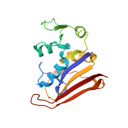
Automated identification of functional dynamic contact networks from X-ray crystallography.

van den Bedem, H.Bhabha, G.Yang, K.Wright, P.E.Fraser, J.S.

(2013) Nat Methods 10: 896-902

  • DOI: https://doi.org/10.1038/nmeth.2592
  • Primary Citation of Related Structures:  
    4KJJ, 4KJK, 4KJL

  • PubMed Abstract: 

    Protein function often depends on the exchange between conformational substates. Allosteric ligand binding or distal mutations can stabilize specific active-site conformations and consequently alter protein function. Observing alternative conformations at low levels of electron density, in addition to comparison of independently determined X-ray crystal structures, can provide mechanistic insights into conformational dynamics. Here we report a new algorithm, CONTACT, that identifies contact networks of conformationally heterogeneous residues directly from high-resolution X-ray crystallography data. Contact networks determined for Escherichia coli dihydrofolate reductase (ecDHFR) predict the observed long-range pattern of NMR chemical shift perturbations of an allosteric mutation. A comparison of contact networks in wild-type and mutant ecDHFR suggests that mutations that alter optimized contact networks of coordinated motions can impair catalytic function. CONTACT-guided mutagenesis can exploit the structure-dynamics-function relationship in protein engineering and design.


  • Organizational Affiliation

    Joint Center for Structural Genomics, Stanford Synchrotron Radiation Lightsource, Stanford, California, USA. vdbedem@slac.stanford.edu


Macromolecules
Find similar proteins by:  (by identity cutoff)  |  3D Structure
Entity ID: 1
MoleculeChains Sequence LengthOrganismDetailsImage
Dihydrofolate reductase159Escherichia coli K-12Mutation(s): 0 
Gene Names: folAUTI89_C0054
EC: 1.5.1.3
UniProt
Find proteins for P0ABQ4 (Escherichia coli (strain K12))
Explore P0ABQ4 
Go to UniProtKB:  P0ABQ4
Entity Groups  
Sequence Clusters30% Identity50% Identity70% Identity90% Identity95% Identity100% Identity
UniProt GroupP0ABQ4
Sequence Annotations
Expand
  • Reference Sequence
Experimental Data & Validation

Experimental Data

  • Method: X-RAY DIFFRACTION
  • Resolution: 1.35 Å
  • R-Value Free: 0.165 
  • R-Value Work: 0.129 
  • R-Value Observed: 0.131 
  • Space Group: P 21 21 21
Unit Cell:
Length ( Å )Angle ( ˚ )
a = 34.32α = 90
b = 45.51β = 90
c = 98.91γ = 90
Software Package:
Software NamePurpose
PHENIXrefinement
PHASERphasing
ELVESrefinement
HKL-2000data reduction
HKL-2000data scaling

Structure Validation

View Full Validation Report


Ligand Structure Quality Assessment 


Entry History 

Deposition Data

Revision History  (Full details and data files)

  • Version 1.0: 2013-08-21
    Type: Initial release
  • Version 1.1: 2013-09-11
    Changes: Database references
  • Version 1.2: 2014-11-12
    Changes: Structure summary
  • Version 1.3: 2017-11-15
    Changes: Refinement description
  • Version 1.4: 2019-07-17
    Changes: Data collection, Refinement description
  • Version 1.5: 2024-02-28
    Changes: Data collection, Database references, Derived calculations