4KI2 | pdb_00004ki2

Crystallographic analysis of an RNA-polymerase sigma-subunit fragment complexed with -10 promoter element ssDNA


Experimental Data Snapshot

  • Method: X-RAY DIFFRACTION
  • Resolution: 3.30 Å
  • R-Value Free: 
    0.262 (Depositor), 0.255 (DCC) 
  • R-Value Work: 
    0.212 (Depositor), 0.216 (DCC) 
  • R-Value Observed: 
    0.216 (Depositor) 

wwPDB Validation 3D Report Full Report

Validation slider image for 4KI2

This is version 1.3 of the entry. See complete history

Literature

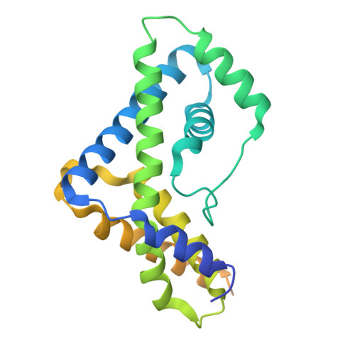
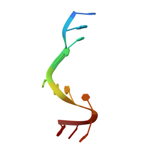
Crystallographic analysis of an RNA polymerase sigma-subunit fragment complexed with -10 promoter element ssDNA: quadruplex formation as a possible tool for engineering crystal contacts in protein-ssDNA complexes.

Feklistov, A.Darst, S.A.

(2013) Acta Crystallogr Sect F Struct Biol Cryst Commun 69: 950-955

  • DOI: https://doi.org/10.1107/S1744309113020368
  • Primary Citation Related Structures: 
    4KI2

  • PubMed Abstract: 

    Structural studies of -10 promoter element recognition by domain 2 of the RNA polymerase σ subunit [Feklistov & Darst (2011), Cell, 147, 1257-1269] reveal an unusual crystal-packing arrangement dominated by G-quartets. The 3'-terminal GGG motif of the oligonucleotide used in crystallization participates in G-quadruplex formation with GGG motifs from symmetry-related complexes. Stacking between neighboring G-quadruplexes results in the formation of pseudo-continuous four-stranded columns running throughout the length of the crystal (G-columns). Here, a new crystal form is presented with a different arrangement of G-columns and it is proposed that the fortuitous finding of G-quartet packing could be useful in engineering crystal contacts in protein-ssDNA complexes.


  • Organizational Affiliation
    • Laboratory of Molecular Biophysics, The Rockefeller University, 1230 York Avenue, New York, NY 10065, USA. afeklistov@rockefeller.edu

Macromolecule Content 

  • Total Structure Weight: 69.93 kDa 
  • Atom Count: 3,798 
  • Modeled Residue Count: 439 
  • Deposited Residue Count: 534 
  • Unique protein chains: 1
  • Unique nucleic acid chains: 1

Macromolecules


Find similar proteins by:|  3D Structure
Entity ID: 2
MoleculeChains  Sequence LengthOrganismDetailsImage
RNA polymerase sigma factorE [auth A],
F [auth B]
245Thermus aquaticusMutation(s): 0 
UniProt
Find proteins for Q9EZJ8 (Thermus aquaticus)
Explore Q9EZJ8 
Go to UniProtKB:  Q9EZJ8
Entity Groups
Sequence Clusters30% Identity50% Identity70% Identity90% Identity95% Identity100% Identity
UniProt GroupQ9EZJ8
Sequence Annotations
Expand
Reference Sequence
Find similar nucleic acids by:  Sequence
Entity ID: 1
MoleculeChains LengthOrganismImage
DNA (5'-D(*TP*GP*TP*AP*CP*AP*AP*TP*GP*GP*G)-3')A [auth D],
B [auth E],
C [auth F],
D [auth G]
11N/A
Sequence Annotations
Expand
Reference Sequence

Experimental Data & Validation

Experimental Data

  • Method: X-RAY DIFFRACTION
  • Resolution: 3.30 Å
  • R-Value Free:  0.262 (Depositor), 0.255 (DCC) 
  • R-Value Work:  0.212 (Depositor), 0.216 (DCC) 
  • R-Value Observed: 0.216 (Depositor) 
Space Group: P 62 2 2
Unit Cell:
Length ( Å )Angle ( ˚ )
a = 118.11α = 90
b = 118.11β = 90
c = 200.845γ = 120
Software Package:
Software NamePurpose
CBASSdata collection
PHASERphasing
PHENIXrefinement
HKL-2000data reduction
HKL-2000data scaling

Structure Validation

View Full Validation Report


Entry History 

Deposition Data

Revision History  (Full details and data files)

  • Version 1.0: 2013-09-04
    Type: Initial release
  • Version 1.1: 2014-09-24
    Changes: Database references
  • Version 1.2: 2018-01-24
    Changes: Structure summary
  • Version 1.3: 2024-02-28
    Changes: Data collection, Database references, Derived calculations