4KFM

Crystal structure of the G protein-gated inward rectifier K+ channel GIRK2 (Kir3.2) in complex with the beta-gamma G protein subunits


Experimental Data Snapshot

  • Method: X-RAY DIFFRACTION
  • Resolution: 3.45 Å
  • R-Value Free: 0.265 
  • R-Value Work: 0.228 
  • R-Value Observed: 0.230 

wwPDB Validation   3D Report Full Report


This is version 1.2 of the entry. See complete history


Literature

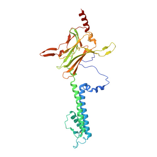
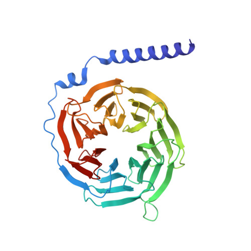
X-ray structure of the mammalian GIRK2-beta gamma G-protein complex.

Whorton, M.R.MacKinnon, R.

(2013) Nature 498: 190-197

  • DOI: https://doi.org/10.1038/nature12241
  • Primary Citation of Related Structures:  
    4KFM

  • PubMed Abstract: 

    G-protein-gated inward rectifier K(+) (GIRK) channels allow neurotransmitters, through G-protein-coupled receptor stimulation, to control cellular electrical excitability. In cardiac and neuronal cells this control regulates heart rate and neural circuit activity, respectively. Here we present the 3.5 Å resolution crystal structure of the mammalian GIRK2 channel in complex with βγ G-protein subunits, the central signalling complex that links G-protein-coupled receptor stimulation to K(+) channel activity. Short-range atomic and long-range electrostatic interactions stabilize four βγ G-protein subunits at the interfaces between four K(+) channel subunits, inducing a pre-open state of the channel. The pre-open state exhibits a conformation that is intermediate between the closed conformation and the open conformation of the constitutively active mutant. The resultant structural picture is compatible with 'membrane delimited' activation of GIRK channels by G proteins and the characteristic burst kinetics of channel gating. The structures also permit a conceptual understanding of how the signalling lipid phosphatidylinositol-4,5-bisphosphate (PIP2) and intracellular Na(+) ions participate in multi-ligand regulation of GIRK channels.


  • Organizational Affiliation

    Laboratory of Molecular Neurobiology and Biophysics, The Rockefeller University, 1230 York Avenue, New York, New York 10065, USA.


Macromolecules
Find similar proteins by:  (by identity cutoff)  |  3D Structure
Entity ID: 1
MoleculeChains Sequence LengthOrganismDetailsImage
G protein-activated inward rectifier potassium channel 2340Mus musculusMutation(s): 0 
Gene Names: Kcnj6
Membrane Entity: Yes 
UniProt & NIH Common Fund Data Resources
Find proteins for P48542 (Mus musculus)
Explore P48542 
Go to UniProtKB:  P48542
IMPC:  MGI:104781
Entity Groups  
Sequence Clusters30% Identity50% Identity70% Identity90% Identity95% Identity100% Identity
UniProt GroupP48542
Sequence Annotations
Expand
  • Reference Sequence
Find similar proteins by:  (by identity cutoff)  |  3D Structure
Entity ID: 2
MoleculeChains Sequence LengthOrganismDetailsImage
Guanine nucleotide-binding protein G(I)/G(S)/G(T) subunit beta-1340Homo sapiensMutation(s): 0 
Gene Names: GNB1
UniProt & NIH Common Fund Data Resources
Find proteins for P62873 (Homo sapiens)
Explore P62873 
Go to UniProtKB:  P62873
PHAROS:  P62873
GTEx:  ENSG00000078369 
Entity Groups  
Sequence Clusters30% Identity50% Identity70% Identity90% Identity95% Identity100% Identity
UniProt GroupP62873
Sequence Annotations
Expand
  • Reference Sequence
Find similar proteins by:  (by identity cutoff)  |  3D Structure
Entity ID: 3
MoleculeChains Sequence LengthOrganismDetailsImage
Guanine nucleotide-binding protein G(I)/G(S)/G(O) subunit gamma-2C [auth G]70Homo sapiensMutation(s): 0 
Gene Names: GNG2
Membrane Entity: Yes 
UniProt & NIH Common Fund Data Resources
Find proteins for P59768 (Homo sapiens)
Explore P59768 
Go to UniProtKB:  P59768
PHAROS:  P59768
GTEx:  ENSG00000186469 
Entity Groups  
Sequence Clusters30% Identity50% Identity70% Identity90% Identity95% Identity100% Identity
UniProt GroupP59768
Sequence Annotations
Expand
  • Reference Sequence
Small Molecules
Ligands 4 Unique
IDChains Name / Formula / InChI Key2D Diagram3D Interactions
PIO
Query on PIO

Download Ideal Coordinates CCD File 
D [auth A][(2R)-2-octanoyloxy-3-[oxidanyl-[(1R,2R,3S,4R,5R,6S)-2,3,6-tris(oxidanyl)-4,5-diphosphonooxy-cyclohexyl]oxy-phosphoryl]oxy-propyl] octanoate
C25 H49 O19 P3
XLNCEHRXXWQMPK-MJUMVPIBSA-N
LMT
Query on LMT

Download Ideal Coordinates CCD File 
K [auth A]DODECYL-BETA-D-MALTOSIDE
C24 H46 O11
NLEBIOOXCVAHBD-QKMCSOCLSA-N
K
Query on K

Download Ideal Coordinates CCD File 
E [auth A],
F [auth A],
G [auth A],
H [auth A],
I [auth A]
POTASSIUM ION
K
NPYPAHLBTDXSSS-UHFFFAOYSA-N
NA
Query on NA

Download Ideal Coordinates CCD File 
J [auth A]SODIUM ION
Na
FKNQFGJONOIPTF-UHFFFAOYSA-N
Experimental Data & Validation

Experimental Data

  • Method: X-RAY DIFFRACTION
  • Resolution: 3.45 Å
  • R-Value Free: 0.265 
  • R-Value Work: 0.228 
  • R-Value Observed: 0.230 
  • Space Group: I 4 2 2
Unit Cell:
Length ( Å )Angle ( ˚ )
a = 127.308α = 90
b = 127.308β = 90
c = 309.392γ = 90
Software Package:
Software NamePurpose
DENZOdata reduction
SCALEPACKdata scaling
PHASERphasing
REFMACrefinement
PDB_EXTRACTdata extraction

Structure Validation

View Full Validation Report


Entry History 

Deposition Data

Revision History  (Full details and data files)

  • Version 1.0: 2013-06-12
    Type: Initial release
  • Version 1.1: 2013-06-19
    Changes: Database references
  • Version 1.2: 2013-09-11
    Changes: Database references