


A new class of type IIA topoisomerase inhibitors with broad-spectrum antibacterial activity
Tari, L.W., Bensen, D.C., Finn, J.To be published.
Experimental Data Snapshot
Starting Model: experimental
View more details
Entity ID: 1 | |||||
|---|---|---|---|---|---|
| Molecule | Chains | Sequence Length | Organism | Details | Image |
| DNA gyrase subunit B | 215 | Escherichia coli K-12 | Mutation(s): 0 Gene Names: acrB, himB, hisU, nalC, parA, pcbA EC: 5.99.1.3 (PDB Primary Data), 5.6.2.2 (UniProt) | 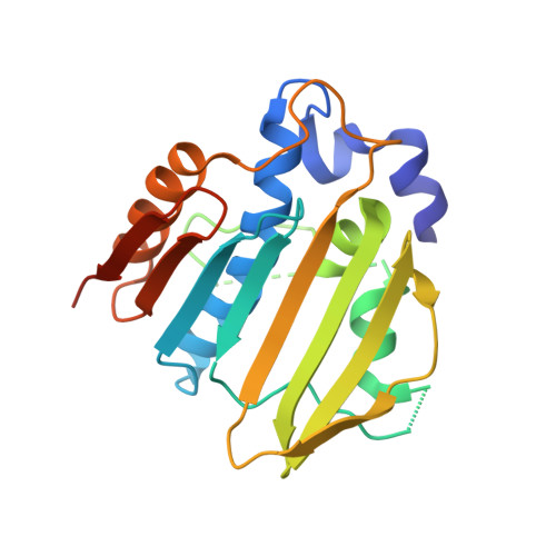 | |
UniProt | |||||
Entity Groups | |||||
| Sequence Clusters | 30% Identity50% Identity70% Identity90% Identity95% Identity100% Identity | ||||
| UniProt Group | P0AES6 | ||||
Sequence AnnotationsExpand | |||||
Reference Sequence | |||||
| Ligands 2 Unique | |||||
|---|---|---|---|---|---|
| ID | Chains | Name / Formula / InChI Key | 2D Diagram | 3D Interactions | |
| DOO Download:Ideal Coordinates CCD File | E [auth A], G [auth B] | 6-fluoro-4-[(3aR,6aR)-hexahydropyrrolo[3,4-b]pyrrol-5(1H)-yl]-N-methyl-2-[(2-methylpyrimidin-5-yl)oxy]-9H-pyrimido[4,5-b]indol-8-amine C22 H23 F N8 O ACMIJDVJWLMBCX-PXAZEXFGSA-N |  | ||
| SO4 Download:Ideal Coordinates CCD File | C [auth A], D [auth A], F [auth B] | SULFATE ION O4 S QAOWNCQODCNURD-UHFFFAOYSA-L |  | ||
| Length ( Å ) | Angle ( ˚ ) |
|---|---|
| a = 47.894 | α = 90 |
| b = 82.477 | β = 100.16 |
| c = 53.74 | γ = 90 |
| Software Name | Purpose |
|---|---|
| REFMAC | refinement |


