4KCF | pdb_00004kcf

X-ray Structure of a KijD3 in Complex with FMN and dTDP-3-amino-2,3,6-trideoxy-4-keto-3-methyl-D-glucose


Experimental Data Snapshot

  • Method: X-RAY DIFFRACTION
  • Resolution: 2.10 Å
  • R-Value Free: 
    0.225 (Depositor), 0.229 (DCC) 
  • R-Value Work: 
    0.175 (Depositor), 0.177 (DCC) 
  • R-Value Observed: 
    0.177 (Depositor) 

Starting Model: experimental
View more details

wwPDB Validation 3D Report Full Report

Validation slider image for 4KCF

Ligand Structure Quality Assessment 


This is version 1.3 of the entry. See complete history

Literature

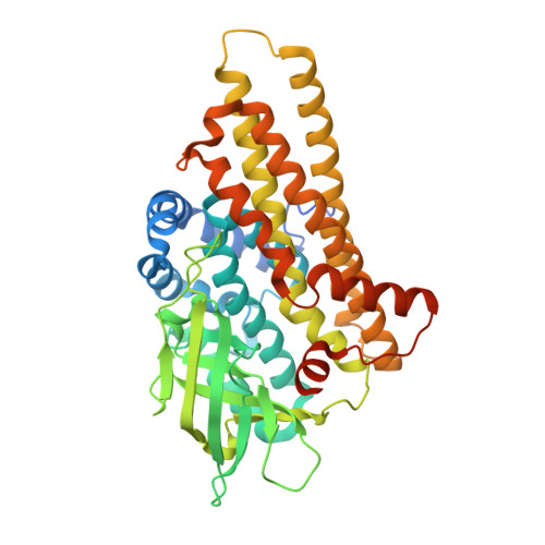
Active site architecture of a sugar N-oxygenase.

Thoden, J.B.Branch, M.C.Zimmer, A.L.Bruender, N.A.Holden, H.M.

(2013) Biochemistry 52: 3191-3193

  • DOI: https://doi.org/10.1021/bi400407x
  • Primary Citation Related Structures: 
    4KCF

  • PubMed Abstract: 

    KijD3 is a flavin-dependent N-oxygenase implicated in the formation of the nitro-containing sugar d-kijanose, found attached to the antibiotic kijanimicin. For this investigation, the structure of KijD3 in complex with FMN and its dTDP-sugar substrate was solved to 2.1 Å resolution. In contrast to the apoenzyme structure, the C-terminus of the protein becomes ordered and projects into the active site cleft [Bruender, N. A., Thoden, J. B., and Holden, H. M. (2010) Biochemistry 49, 3517-3524]. The amino group of the dTDP-aminosugar that is oxidized is located 4.9 Å from C4a of the flavin ring. The model provides a molecular basis for understanding the manner in which KijD3 catalyzes its unusual chemical transformation.


  • Organizational Affiliation
    • Department of Biochemistry, University of Wisconsin , Madison, Wisconsin 53706, United States.

Macromolecule Content 

  • Total Structure Weight: 49.35 kDa 
  • Atom Count: 3,199 
  • Modeled Residue Count: 407 
  • Deposited Residue Count: 454 
  • Unique protein chains: 1

Macromolecules

Find similar proteins by:|  3D Structure
Entity ID: 1
MoleculeChains  Sequence LengthOrganismDetailsImage
FAD-dependent oxidoreductase454Actinomadura kijaniataMutation(s): 0 
Gene Names: KijD3
UniProt
Find proteins for B3TMR1 (Actinomadura kijaniata)
Explore B3TMR1 
Go to UniProtKB:  B3TMR1
Entity Groups
Sequence Clusters30% Identity50% Identity70% Identity90% Identity95% Identity100% Identity
UniProt GroupB3TMR1
Sequence Annotations
Expand
Reference Sequence

Small Molecules

Ligands 2 Unique
IDChains Name / Formula / InChI Key2D Diagram3D Interactions
AKM

Query on AKM


Download:Ideal Coordinates CCD File
B [auth A][(2R,4S,6R)-4-azanyl-4,6-dimethyl-5,5-bis(oxidanyl)oxan-2-yl] [[(2R,3S,5R)-5-[5-methyl-2,4-bis(oxidanylidene)pyrimidin-1-yl]-3-oxidanyl-oxolan-2-yl]methoxy-oxidanyl-phosphoryl] hydrogen phosphate
C17 H29 N3 O14 P2
GACNZVKWDHTCBB-HHMBNNFFSA-N
FMN

Query on FMN


Download:Ideal Coordinates CCD File
C [auth A]FLAVIN MONONUCLEOTIDE
C17 H21 N4 O9 P
FVTCRASFADXXNN-SCRDCRAPSA-N

Experimental Data & Validation

Experimental Data

  • Method: X-RAY DIFFRACTION
  • Resolution: 2.10 Å
  • R-Value Free:  0.225 (Depositor), 0.229 (DCC) 
  • R-Value Work:  0.175 (Depositor), 0.177 (DCC) 
  • R-Value Observed: 0.177 (Depositor) 
Space Group: I 2 2 2
Unit Cell:
Length ( Å )Angle ( ˚ )
a = 70.828α = 90
b = 81.297β = 90
c = 152.628γ = 90
Software Package:
Software NamePurpose
HKL-3000data collection
PHASERphasing
REFMACrefinement
HKL-3000data reduction
HKL-3000data scaling

Structure Validation

View Full Validation Report


Ligand Structure Quality Assessment 


Entry History 

Deposition Data

Revision History  (Full details and data files)

  • Version 1.0: 2013-05-22
    Type: Initial release
  • Version 1.1: 2013-05-29
    Changes: Atomic model, Non-polymer description
  • Version 1.2: 2013-12-18
    Changes: Database references
  • Version 1.3: 2023-09-20
    Changes: Data collection, Database references, Derived calculations, Refinement description