4KAB | pdb_00004kab

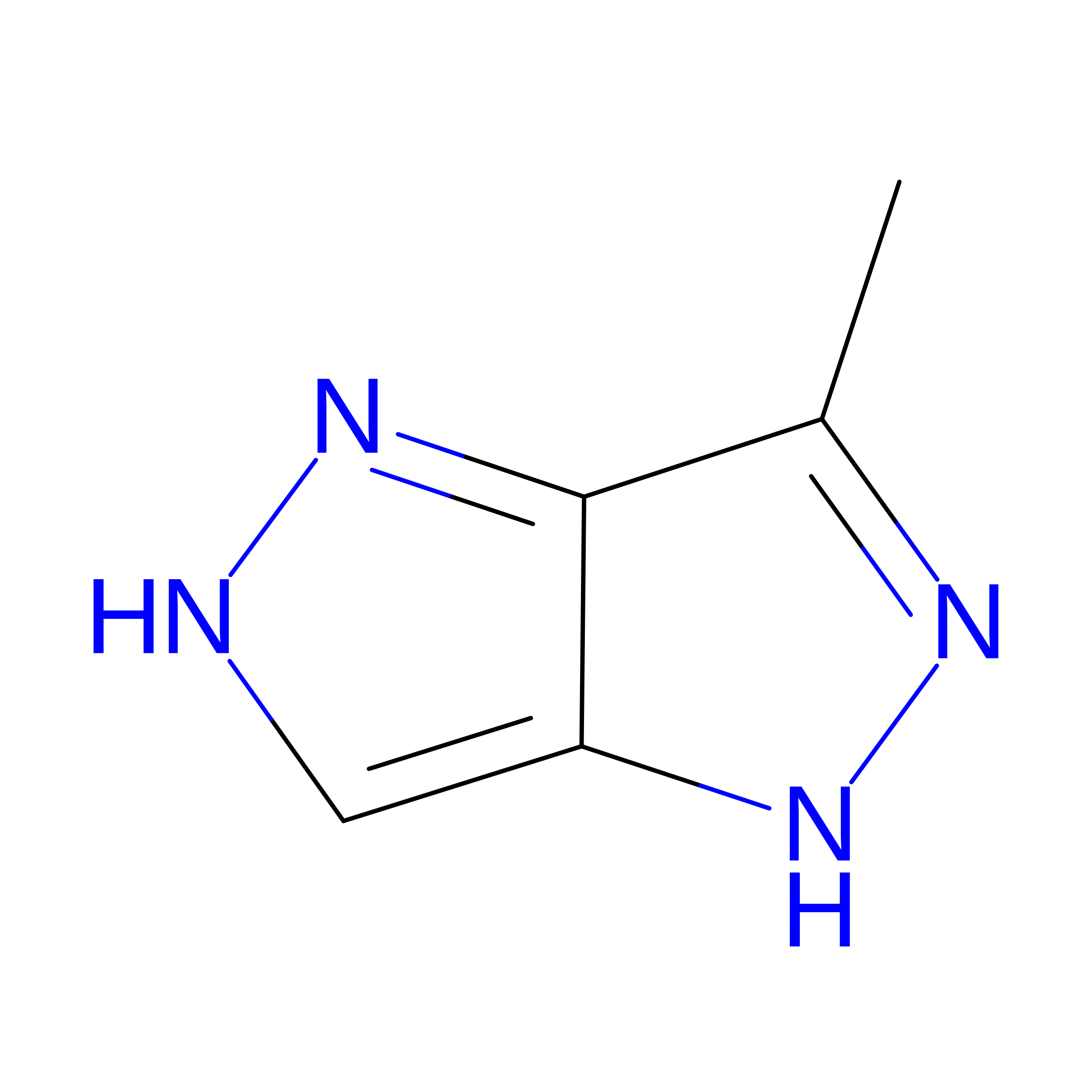
FOCAL ADHESION KINASE CATALYTIC DOMAIN IN COMPLEX WITH 3-Methyl-1,4-dihydro-pyrazolo[4,5-c]pyrazole


Experimental Data Snapshot

  • Method: X-RAY DIFFRACTION
  • Resolution: 2.71 Å
  • R-Value Free: 
    0.219 (Depositor), 0.227 (DCC) 
  • R-Value Work: 
    0.171 (Depositor), 0.178 (DCC) 
  • R-Value Observed: 
    0.173 (Depositor) 

wwPDB Validation 3D Report Full Report

Validation slider image for 4KAB

This is version 1.2 of the entry. See complete history. 

Literature

Fragment-based discovery of focal adhesion kinase inhibitors.

Gradler, U., Bomke, J., Musil, D., Dresing, V., Lehmann, M., Holzemann, G., Greiner, H., Esdar, C., Krier, M., Heinrich, T.

(2013) Bioorg Med Chem Lett 23: 5401-5409

  • DOI: https://doi.org/10.1016/j.bmcl.2013.07.050
  • Primary Citation Related Structures: 
    4K8A, 4K9Y, 4KAB, 4KAO

  • PubMed Abstract: 

    Chemically diverse fragment hits of focal adhesion kinase (FAK) were discovered by surface plasmon resonance (SPR) screening of our in-house fragment library. Site specific binding of the primary hits was confirmed in a competition setup using a high-affinity ATP-site inhibitor of FAK. Protein crystallography revealed the binding mode of 41 out of 48 selected fragment hits within the ATP-site. Structural comparison of the fragment binding modes with a DFG-out inhibitor of FAK initiated first synthetic follow-up optimization leading to improved binding affinity.


  • Organizational Affiliation: 
    • Merck KGaA, Merck Serono Research, Darmstadt, Germany. ulrich.graedler@merckgroup.com

Macromolecule Content 

  • Total Structure Weight: 64.16 kDa 
  • Atom Count: 4,221 
  • Modeled Residue Count: 513 
  • Deposited Residue Count: 558 
  • Unique protein chains: 1

Macromolecules

Find similar proteins by:|  3D Structure
Entity ID: 1
MoleculeChains  Sequence LengthOrganismDetailsImage
Focal adhesion kinase 1
A, B
279Homo sapiensMutation(s): 0 
Gene Names: PTK2, FAK, FAK1
EC: 2.7.10.2
UniProt & NIH Common Fund Data Resources
Find proteins for Q05397 (Homo sapiens)
Explore Q05397 
Go to UniProtKB:  Q05397
PHAROS:  Q05397
GTEx:  ENSG00000169398 
Entity Groups
Sequence Clusters30% Identity50% Identity70% Identity90% Identity95% Identity100% Identity
UniProt GroupQ05397
Sequence Annotations
Expand
Reference Sequence

Experimental Data & Validation

Experimental Data

  • Method: X-RAY DIFFRACTION
  • Resolution: 2.71 Å
  • R-Value Free:  0.219 (Depositor), 0.227 (DCC) 
  • R-Value Work:  0.171 (Depositor), 0.178 (DCC) 
  • R-Value Observed: 0.173 (Depositor) 
Space Group: P 21 21 21
Unit Cell:
Length ( Å )Angle ( ˚ )
a = 45.96α = 90
b = 88.59β = 90
c = 136.89γ = 90
Software Package:
Software NamePurpose
PHASERphasing
BUSTERrefinement
XDSdata reduction
XSCALEdata scaling

Structure Validation

View Full Validation Report


Entry History 

Deposition Data

Revision History  (Full details and data files)

  • Version 1.0: 2013-09-11
    Type: Initial release
  • Version 1.1: 2013-10-09
    Changes: Database references
  • Version 1.2: 2024-11-27
    Changes: Data collection, Database references, Derived calculations, Structure summary