4K5Z | pdb_00004k5z

Crystal Structure of Human Chymase in Complex with Fragment Inhibitor 6-chloro-2,3-dihydro-1H-isoindol-1-one


Experimental Data Snapshot

  • Method: X-RAY DIFFRACTION
  • Resolution: 1.80 Å
  • R-Value Free: 
    0.263 (Depositor), 0.267 (DCC) 
  • R-Value Work: 
    0.223 (Depositor), 0.226 (DCC) 
  • R-Value Observed: 
    0.225 (Depositor) 

wwPDB Validation 3D Report Full Report

Validation slider image for 4K5Z

Ligand Structure Quality Assessment 


This is version 1.3 of the entry. See complete history

Literature

Discovery of Potent, Selective Chymase Inhibitors via Fragment Linking Strategies.

Taylor, S.J.Padyana, A.K.Abeywardane, A.Liang, S.Hao, M.H.De Lombaert, S.Proudfoot, J.Farmer, B.S.Li, X.Collins, B.Martin, L.Albaugh, D.R.Hill-Drzewi, M.Pullen, S.S.Takahashi, H.

(2013) J Med Chem 56: 4465-4481

  • DOI: https://doi.org/10.1021/jm400138z
  • Primary Citation Related Structures: 
    4K2Y, 4K5Z, 4K60, 4K69

  • PubMed Abstract: 

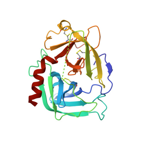
    Chymase plays an important and diverse role in the homeostasis of a number of cardiovascular processes. Herein, we describe the identification of potent, selective chymase inhibitors, developed using fragment-based, structure-guided linking and optimization techniques. High-concentration biophysical screening methods followed by high-throughput crystallography identified an oxindole fragment bound to the S1 pocket of the protein exhibiting a novel interaction pattern hitherto not observed in chymase inhibitors. X-ray crystallographic structures were used to guide the elaboration/linking of the fragment, ultimately leading to a potent inhibitor that was >100-fold selective over cathepsin G and that mitigated a number of liabilities associated with poor physicochemical properties of the series it was derived from.


  • Organizational Affiliation
    • Department of Medicinal Chemistry, Boehringer Ingelheim Pharmaceuticals Inc., 900 Ridgebury Road, Ridgefield, Connecticut 06877-0368, USA. steven.taylor@boehringeringelheim.com

Macromolecule Content 

  • Total Structure Weight: 25.45 kDa 
  • Atom Count: 1,968 
  • Modeled Residue Count: 220 
  • Deposited Residue Count: 226 
  • Unique protein chains: 1

Macromolecules

Find similar proteins by:|  3D Structure
Entity ID: 1
MoleculeChains  Sequence LengthOrganismDetailsImage
Chymase226Homo sapiensMutation(s): 0 
Gene Names: CMA1CYHCYM
EC: 3.4.21.39
UniProt & NIH Common Fund Data Resources
Find proteins for P23946 (Homo sapiens)
Explore P23946 
Go to UniProtKB:  P23946
PHAROS:  P23946
GTEx:  ENSG00000092009 
Entity Groups
Sequence Clusters30% Identity50% Identity70% Identity90% Identity95% Identity100% Identity
UniProt GroupP23946
Glycosylation
Glycosylation Sites: 1Go to GlyGen: P23946-1
Sequence Annotations
Expand
Reference Sequence

Small Molecules

Binding Affinity Annotations 
IDSourceBinding Affinity
1P7 BindingDB:  4K5Z IC50: 1.00e+6 (nM) from 1 assay(s)

Experimental Data & Validation

Experimental Data

  • Method: X-RAY DIFFRACTION
  • Resolution: 1.80 Å
  • R-Value Free:  0.263 (Depositor), 0.267 (DCC) 
  • R-Value Work:  0.223 (Depositor), 0.226 (DCC) 
  • R-Value Observed: 0.225 (Depositor) 
Space Group: P 43
Unit Cell:
Length ( Å )Angle ( ˚ )
a = 74.609α = 90
b = 74.609β = 90
c = 49.897γ = 90
Software Package:
Software NamePurpose
d*TREKdata reduction
PHENIXrefinement
PDB_EXTRACTdata extraction
StructureStudiodata collection
d*TREKdata scaling
CNXphasing

Structure Validation

View Full Validation Report


Ligand Structure Quality Assessment 


Entry History 

Deposition Data

Revision History  (Full details and data files)

  • Version 1.0: 2013-05-29
    Type: Initial release
  • Version 1.1: 2013-07-03
    Changes: Database references
  • Version 1.2: 2020-07-29
    Type: Remediation
    Reason: Carbohydrate remediation
    Changes: Data collection, Database references, Derived calculations, Structure summary
  • Version 1.3: 2024-11-20
    Changes: Data collection, Database references, Structure summary