4K40 | pdb_00004k40

Peptidoglycan O-acetylesterase in action, 0 min


Experimental Data Snapshot

  • Method: X-RAY DIFFRACTION
  • Resolution: 2.63 Å
  • R-Value Free: 
    0.238 (Depositor), 0.240 (DCC) 
  • R-Value Work: 
    0.165 (Depositor), 0.172 (DCC) 
  • R-Value Observed: 
    0.169 (Depositor) 

wwPDB Validation 3D Report Full Report

Validation slider image for 4K40

This is version 1.3 of the entry. See complete history

Literature

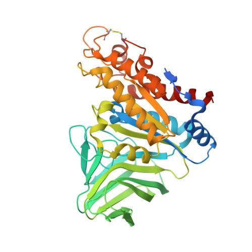
Visualization of a substrate-induced productive conformation of the catalytic triad of the Neisseria meningitidis peptidoglycan O-acetylesterase reveals mechanistic conservation in SGNH esterase family members.

Williams, A.H.Veyrier, F.J.Bonis, M.Michaud, Y.Raynal, B.Taha, M.K.White, S.W.Haouz, A.Boneca, I.G.

(2014) Acta Crystallogr D Biol Crystallogr 70: 2631-2639

  • DOI: https://doi.org/10.1107/S1399004714016770
  • Primary Citation Related Structures: 
    4K3U, 4K40, 4K7J, 4K9S

  • PubMed Abstract: 

    Peptidoglycan O-acetylesterase (Ape1), which is required for host survival in Neisseria sp., belongs to the diverse SGNH hydrolase superfamily, which includes important viral and bacterial virulence factors. Here, multi-domain crystal structures of Ape1 with an SGNH catalytic domain and a newly identified putative peptidoglycan-detection module are reported. Enzyme catalysis was performed in Ape1 crystals and key catalytic intermediates along the SGNH esterase hydrolysis reaction pathway were visualized, revealing a substrate-induced productive conformation of the catalytic triad, a mechanistic detail that has not previously been observed. This substrate-induced productive conformation of the catalytic triad shifts the established dogma on these enzymes, generating valuable insight into the structure-based design of drugs targeting the SGNH esterase superfamily.


  • Organizational Affiliation
    • Institut Pasteur, Unité Biologie et génétique de la paroi bactérienne, Dept. Microbiologie, 28 Rue du Dr. Roux, 75015 Paris, France.

Macromolecule Content 

  • Total Structure Weight: 81.58 kDa 
  • Atom Count: 5,386 
  • Modeled Residue Count: 698 
  • Deposited Residue Count: 758 
  • Unique protein chains: 1

Macromolecules

Find similar proteins by:|  3D Structure
Entity ID: 1
MoleculeChains  Sequence LengthOrganismDetailsImage
GDSL-like Lipase/Acylhydrolase family protein
A, B
379Neisseria meningitidis LNP21362Mutation(s): 0 
Gene Names: N875_00620

Experimental Data & Validation

Experimental Data

  • Method: X-RAY DIFFRACTION
  • Resolution: 2.63 Å
  • R-Value Free:  0.238 (Depositor), 0.240 (DCC) 
  • R-Value Work:  0.165 (Depositor), 0.172 (DCC) 
  • R-Value Observed: 0.169 (Depositor) 
Space Group: P 21 21 21
Unit Cell:
Length ( Å )Angle ( ˚ )
a = 71.872α = 90
b = 79.493β = 90
c = 124.383γ = 90
Software Package:
Software NamePurpose
MxCuBEdata collection
PHENIXmodel building
PHENIXrefinement
XDSdata reduction
XDSdata scaling
PHENIXphasing

Structure Validation

View Full Validation Report


Entry History 

Deposition Data

Revision History  (Full details and data files)

  • Version 1.0: 2014-09-03
    Type: Initial release
  • Version 1.1: 2014-10-22
    Changes: Database references
  • Version 1.2: 2017-06-21
    Changes: Database references, Source and taxonomy
  • Version 1.3: 2024-10-30
    Changes: Data collection, Database references, Structure summary