


Crystal structure of D-erythrulose 4-phosphate dehydrogenase from Brucella melitensis, solved by iodide SAD
Seattle Structural Genomics Center for Infectious Disease (SSGCID), Abendroth, J., Clifton, M.C., Lorimer, D., Edwards, T.E.To be published.
Experimental Data Snapshot
wwPDB Validation 3D Report Full Report

Entity ID: 1 | |||||
|---|---|---|---|---|---|
| Molecule | Chains | Sequence Length | Organism | Details | Image |
| D-erythrulose 4-phosphate dehydrogenase | 331 | Brucella melitensis bv. 1 str. 16M | Mutation(s): 0 Gene Names: BAWG_2138, BMEII0428 EC: 1.1.1 | 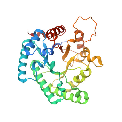 | |
UniProt | |||||
Entity Groups | |||||
| Sequence Clusters | 30% Identity50% Identity70% Identity90% Identity95% Identity100% Identity | ||||
| UniProt Group | Q8YCV0 | ||||
Sequence AnnotationsExpand | |||||
Reference Sequence | |||||
| Ligands 2 Unique | |||||
|---|---|---|---|---|---|
| ID | Chains | Name / Formula / InChI Key | 2D Diagram | 3D Interactions | |
| GOL Download:Ideal Coordinates CCD File | C [auth A] | GLYCEROL C3 H8 O3 PEDCQBHIVMGVHV-UHFFFAOYSA-N |  | ||
| EDO Download:Ideal Coordinates CCD File | D [auth A], E [auth B], F [auth B], G [auth B], H [auth B] | 1,2-ETHANEDIOL C2 H6 O2 LYCAIKOWRPUZTN-UHFFFAOYSA-N |  | ||
| Length ( Å ) | Angle ( ˚ ) |
|---|---|
| a = 103.5 | α = 90 |
| b = 103.5 | β = 90 |
| c = 256.54 | γ = 120 |
| Software Name | Purpose |
|---|---|
| XSCALE | data scaling |
| PHASER | phasing |
| REFMAC | refinement |
| PDB_EXTRACT | data extraction |
| JDirector | data collection |
| XDS | data reduction |


