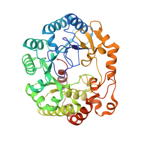
4K36

His6 tagged anSMEcpe with bound AdoMet


Experimental Data Snapshot

  • Method: X-RAY DIFFRACTION
  • Resolution: 1.62 Å
  • R-Value Free: 0.195 
  • R-Value Work: 0.182 
  • R-Value Observed: 0.183 

wwPDB Validation   3D Report Full Report


Ligand Structure Quality Assessment 


This is version 1.2 of the entry. See complete history


Literature

X-ray structure of an AdoMet radical activase reveals an anaerobic solution for formylglycine posttranslational modification.

Goldman, P.J.Grove, T.L.Sites, L.A.McLaughlin, M.I.Booker, S.J.Drennan, C.L.

(2013) Proc Natl Acad Sci U S A 110: 8519-8524

  • DOI: https://doi.org/10.1073/pnas.1302417110
  • Primary Citation of Related Structures:  
    4K36, 4K37, 4K38, 4K39

  • PubMed Abstract: 

    Arylsulfatases require a maturating enzyme to perform a co- or posttranslational modification to form a catalytically essential formylglycine (FGly) residue. In organisms that live aerobically, molecular oxygen is used enzymatically to oxidize cysteine to FGly. Under anaerobic conditions, S-adenosylmethionine (AdoMet) radical chemistry is used. Here we present the structures of an anaerobic sulfatase maturating enzyme (anSME), both with and without peptidyl-substrates, at 1.6-1.8 Å resolution. We find that anSMEs differ from their aerobic counterparts in using backbone-based hydrogen-bonding patterns to interact with their peptidyl-substrates, leading to decreased sequence specificity. These anSME structures from Clostridium perfringens are also the first of an AdoMet radical enzyme that performs dehydrogenase chemistry. Together with accompanying mutagenesis data, a mechanistic proposal is put forth for how AdoMet radical chemistry is coopted to perform a dehydrogenation reaction. In the oxidation of cysteine or serine to FGly by anSME, we identify D277 and an auxiliary [4Fe-4S] cluster as the likely acceptor of the final proton and electron, respectively. D277 and both auxiliary clusters are housed in a cysteine-rich C-terminal domain, termed SPASM domain, that contains homology to ~1,400 other unique AdoMet radical enzymes proposed to use [4Fe-4S] clusters to ligate peptidyl-substrates for subsequent modification. In contrast to this proposal, we find that neither auxiliary cluster in anSME bind substrate, and both are fully ligated by cysteine residues. Instead, our structural data suggest that the placement of these auxiliary clusters creates a conduit for electrons to travel from the buried substrate to the protein surface.


  • Organizational Affiliation

    Department of Chemistry and Biology, Massachusetts Institute of Technology, Cambridge, MA 02139, USA.


Macromolecules
Find similar proteins by:  (by identity cutoff)  |  3D Structure
Entity ID: 1
MoleculeChains Sequence LengthOrganismDetailsImage
Anaerobic sulfatase-maturating enzyme
A, B
392Clostridium perfringens ATCC 13124Mutation(s): 0 
Gene Names: CPF_0616
EC: 1.8.98 (PDB Primary Data), 1.8.98.7 (UniProt)
UniProt
Find proteins for Q0TTH1 (Clostridium perfringens (strain ATCC 13124 / DSM 756 / JCM 1290 / NCIMB 6125 / NCTC 8237 / Type A))
Explore Q0TTH1 
Go to UniProtKB:  Q0TTH1
Entity Groups  
Sequence Clusters30% Identity50% Identity70% Identity90% Identity95% Identity100% Identity
UniProt GroupQ0TTH1
Sequence Annotations
Expand
  • Reference Sequence
Small Molecules
Ligands 3 Unique
IDChains Name / Formula / InChI Key2D Diagram3D Interactions
SAM
Query on SAM

Download Ideal Coordinates CCD File 
F [auth A],
K [auth B]
S-ADENOSYLMETHIONINE
C15 H22 N6 O5 S
MEFKEPWMEQBLKI-FCKMPRQPSA-N
SF4
Query on SF4

Download Ideal Coordinates CCD File 
C [auth A]
D [auth A]
E [auth A]
H [auth B]
I [auth B]
C [auth A],
D [auth A],
E [auth A],
H [auth B],
I [auth B],
J [auth B]
IRON/SULFUR CLUSTER
Fe4 S4
LJBDFODJNLIPKO-UHFFFAOYSA-N
CL
Query on CL

Download Ideal Coordinates CCD File 
G [auth A],
L [auth B],
M [auth B]
CHLORIDE ION
Cl
VEXZGXHMUGYJMC-UHFFFAOYSA-M
Experimental Data & Validation

Experimental Data

  • Method: X-RAY DIFFRACTION
  • Resolution: 1.62 Å
  • R-Value Free: 0.195 
  • R-Value Work: 0.182 
  • R-Value Observed: 0.183 
  • Space Group: P 1 21 1
Unit Cell:
Length ( Å )Angle ( ˚ )
a = 46.561α = 90
b = 92.617β = 94
c = 92.348γ = 90
Software Package:
Software NamePurpose
PHENIXrefinement
PDB_EXTRACTdata extraction
APEXdata collection
HKL-2000data reduction
HKL-2000data scaling
PHASERphasing

Structure Validation

View Full Validation Report


Ligand Structure Quality Assessment 


Entry History 

Deposition Data

Revision History  (Full details and data files)

  • Version 1.0: 2013-05-08
    Type: Initial release
  • Version 1.1: 2013-06-12
    Changes: Database references
  • Version 1.2: 2024-02-28
    Changes: Data collection, Database references, Derived calculations