4K2S | pdb_00004k2s

Crystal structure of the mutant P317A of d-mannonate dehydratase from chromohalobacter salexigens complexed with mg and d-gluconate


Experimental Data Snapshot

  • Method: X-RAY DIFFRACTION
  • Resolution: 1.70 Å
  • R-Value Free: 
    0.203 (Depositor), 0.203 (DCC) 
  • R-Value Work: 
    0.169 (Depositor), 0.169 (DCC) 
  • R-Value Observed: 
    0.170 (Depositor) 

Starting Model: experimental
View more details

wwPDB Validation 3D Report Full Report

Validation slider image for 4K2S

Ligand Structure Quality Assessment 


This is version 2.1 of the entry. See complete history

Literature

Crystal structure of the mutant P317A of d-mannonate dehydratase from chromohalobacter salexigens complexed with mg and d-gluconate

Fedorov, A.A.Fedorov, E.V.Wichelecki, D.Gerlt, J.A.Almo, S.C.

To be published.

Macromolecule Content 

  • Total Structure Weight: 365.73 kDa 
  • Atom Count: 28,312 
  • Modeled Residue Count: 3,136 
  • Deposited Residue Count: 3,240 
  • Unique protein chains: 1

Macromolecules

Find similar proteins by:|  3D Structure
Entity ID: 1
MoleculeChains  Sequence LengthOrganismDetailsImage
D-mannonate dehydratase
A, B, C, D, E
A, B, C, D, E, F, G, H
405Chromohalobacter israelensis DSM 3043Mutation(s): 1 
Gene Names: Csal_2974
EC: 4.2.1.8 (PDB Primary Data), 4.2.1 (UniProt), 4.2.1.39 (UniProt)
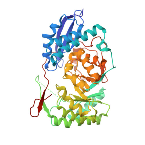
UniProt
Find proteins for Q1QT89 (Chromohalobacter israelensis (strain ATCC BAA-138 / DSM 3043 / CIP 106854 / NCIMB 13768 / 1H11))
Explore Q1QT89 
Go to UniProtKB:  Q1QT89
Entity Groups
Sequence Clusters30% Identity50% Identity70% Identity90% Identity95% Identity100% Identity
UniProt GroupQ1QT89
Sequence Annotations
Expand
Reference Sequence

Small Molecules

Ligands 4 Unique
IDChains Name / Formula / InChI Key2D Diagram3D Interactions
GCO

Query on GCO


Download:Ideal Coordinates CCD File
BA [auth F]
FA [auth G]
I [auth A]
IA [auth H]
L [auth B]
BA [auth F],
FA [auth G],
I [auth A],
IA [auth H],
L [auth B],
O [auth C],
S [auth D],
W [auth E]
D-gluconic acid
C6 H12 O7
RGHNJXZEOKUKBD-SQOUGZDYSA-N
GOL

Query on GOL


Download:Ideal Coordinates CCD File
R [auth C]GLYCEROL
C3 H8 O3
PEDCQBHIVMGVHV-UHFFFAOYSA-N
CL

Query on CL


Download:Ideal Coordinates CCD File
AA [auth E]
DA [auth F]
EA [auth F]
K [auth A]
V [auth D]
AA [auth E],
DA [auth F],
EA [auth F],
K [auth A],
V [auth D],
Z [auth E]
CHLORIDE ION
Cl
VEXZGXHMUGYJMC-UHFFFAOYSA-M
MG

Query on MG


Download:Ideal Coordinates CCD File
CA [auth F]
GA [auth G]
HA [auth G]
J [auth A]
JA [auth H]
CA [auth F],
GA [auth G],
HA [auth G],
J [auth A],
JA [auth H],
M [auth B],
N [auth B],
P [auth C],
Q [auth C],
T [auth D],
U [auth D],
X [auth E],
Y [auth E]
MAGNESIUM ION
Mg
JLVVSXFLKOJNIY-UHFFFAOYSA-N

Experimental Data & Validation

Experimental Data

  • Method: X-RAY DIFFRACTION
  • Resolution: 1.70 Å
  • R-Value Free:  0.203 (Depositor), 0.203 (DCC) 
  • R-Value Work:  0.169 (Depositor), 0.169 (DCC) 
  • R-Value Observed: 0.170 (Depositor) 
Space Group: C 1 2 1
Unit Cell:
Length ( Å )Angle ( ˚ )
a = 196.334α = 90
b = 85.79β = 110.47
c = 195.638γ = 90
Software Package:
Software NamePurpose
PHENIXrefinement

Structure Validation

View Full Validation Report


Ligand Structure Quality Assessment 


Entry History 

Deposition Data

Revision History  (Full details and data files)

  • Version 1.0: 2014-04-09
    Type: Initial release
  • Version 2.0: 2020-07-29
    Type: Remediation
    Reason: Carbohydrate remediation
    Changes: Atomic model, Database references, Derived calculations, Structure summary
  • Version 2.1: 2023-09-20
    Changes: Data collection, Database references, Refinement description, Structure summary