4K1L | pdb_00004k1l

4,4-Dioxo-5,6-dihydro-[1,4,3]oxathiazines, a novel class of 11 beta-HSD1 inhibitors for the treatment of diabetes


Experimental Data Snapshot

  • Method: X-RAY DIFFRACTION
  • Resolution: 1.96 Å
  • R-Value Free: 
    0.202 (Depositor), 0.212 (DCC) 
  • R-Value Work: 
    0.167 (Depositor), 0.173 (DCC) 
  • R-Value Observed: 
    0.169 (Depositor) 

Starting Model: experimental
View more details

wwPDB Validation 3D Report Full Report

Validation slider image for 4K1L

Ligand Structure Quality Assessment 


This is version 1.1 of the entry. See complete history

Literature

1,1-Dioxo-5,6-dihydro-[4,1,2]oxathiazines, a novel class of 11-HSD1 inhibitors for the treatment of diabetes.

Bohme, T.Engel, C.K.Farjot, G.Gussregen, S.Haack, T.Tschank, G.Ritter, K.

(2013) Bioorg Med Chem Lett 23: 4685-4691

  • DOI: https://doi.org/10.1016/j.bmcl.2013.05.102
  • Primary Citation Related Structures: 
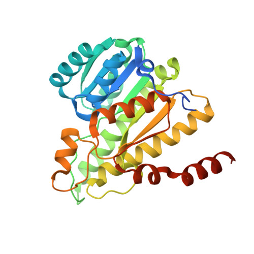
    4K1L, 4K26

  • PubMed Abstract: 

    Racemic cis-1,1-dioxo-5,6-dihydro-[4,1,2]oxathiazine derivative 4a was isolated as an impurity in a sample of a hit from a HTS campaign on 11β-hydroxysteroid dehydrogenase type 1 (11β-HSD1). After separation by chiral chromatography the 4a-S, 8a-R enantiomer of compound 4a was identified as the true, potent enzyme inhibitor. The cocrystal structure of 4a with human and murine 11ß-HSD1 revealed the unique binding mode of the oxathiazine series. SAR elucidation and optimization in regard to metabolic stability led to monocyclic tetramethyloxathiazines as exemplified by compound 21g.


  • Organizational Affiliation
    • Sanofi Deutschland GmbH, R&D, Industriepark Höchst, 65926 Frankfurt am Main, Germany.

Macromolecule Content 

  • Total Structure Weight: 131.47 kDa 
  • Atom Count: 9,120 
  • Modeled Residue Count: 1,056 
  • Deposited Residue Count: 1,144 
  • Unique protein chains: 1

Macromolecules

Find similar proteins by:|  3D Structure
Entity ID: 1
MoleculeChains  Sequence LengthOrganismDetailsImage
Corticosteroid 11-beta-dehydrogenase isozyme 1
A, B, C, D
286Homo sapiensMutation(s): 1 
Gene Names: HSD11B1HSD11HSD11L
EC: 1.1.1.146 (PDB Primary Data), 1.1.1.201 (UniProt)
UniProt & NIH Common Fund Data Resources
Find proteins for P28845 (Homo sapiens)
Explore P28845 
Go to UniProtKB:  P28845
PHAROS:  P28845
GTEx:  ENSG00000117594 
Entity Groups
Sequence Clusters30% Identity50% Identity70% Identity90% Identity95% Identity100% Identity
UniProt GroupP28845
Sequence Annotations
Expand
Reference Sequence

Small Molecules

Ligands 2 Unique
IDChains Name / Formula / InChI Key2D Diagram3D Interactions
NDP

Query on NDP


Download:Ideal Coordinates CCD File
F [auth A],
H [auth B],
J [auth C],
L [auth D]
NADPH DIHYDRO-NICOTINAMIDE-ADENINE-DINUCLEOTIDE PHOSPHATE
C21 H30 N7 O17 P3
ACFIXJIJDZMPPO-NNYOXOHSSA-N
SFF

Query on SFF


Download:Ideal Coordinates CCD File
E [auth A],
G [auth B],
I [auth C],
K [auth D]
(4aS,8aR)-N-cyclohexyl-4a,5,6,7,8,8a-hexahydro-4,1,2-benzoxathiazin-3-amine 1,1-dioxide
C13 H22 N2 O3 S
ZMDVQNCMHSXAPA-NWDGAFQWSA-N

Experimental Data & Validation

Experimental Data

  • Method: X-RAY DIFFRACTION
  • Resolution: 1.96 Å
  • R-Value Free:  0.202 (Depositor), 0.212 (DCC) 
  • R-Value Work:  0.167 (Depositor), 0.173 (DCC) 
  • R-Value Observed: 0.169 (Depositor) 
Space Group: P 1 21 1
Unit Cell:
Length ( Å )Angle ( ˚ )
a = 57.361α = 90
b = 150.941β = 93.79
c = 74.169γ = 90
Software Package:
Software NamePurpose
BUSTERrefinement
XDSdata reduction
XSCALEdata scaling
BUSTERphasing

Structure Validation

View Full Validation Report


Ligand Structure Quality Assessment 


Entry History 

Deposition Data

Revision History  (Full details and data files)

  • Version 1.0: 2014-04-09
    Type: Initial release
  • Version 1.1: 2023-09-20
    Changes: Data collection, Database references, Derived calculations, Refinement description