CARMIL leading edge localization depends on a non-canonical PH domain and dimerization.
Zwolak, A., Yang, C., Feeser, E.A., Michael Ostap, E., Svitkina, T., Dominguez, R.(2013) Nat Commun 4: 2523-2523
- PubMed: 24071777 Search on PubMedSearch on PubMed Central
- DOI: https://doi.org/10.1038/ncomms3523
- Primary Citation Related Structures:
4K17 - PubMed Abstract:
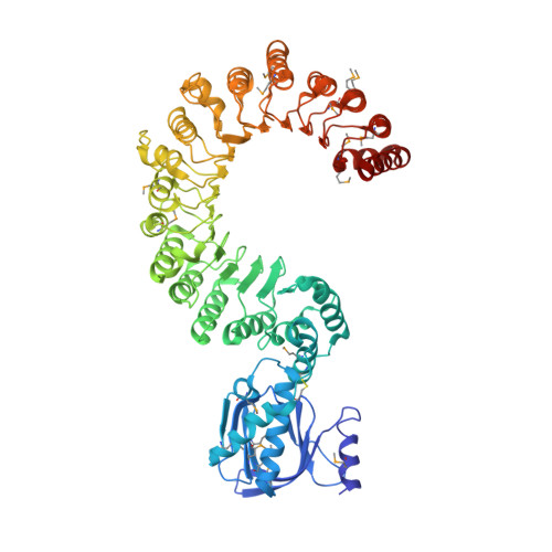
CARMIL is an approximately 1,370-amino-acid cytoskeletal scaffold that has crucial roles in cell motility and tissue development through interactions with cytoskeletal effectors and regulation of capping protein at the leading edge. However, the mechanism of CARMIL leading edge localization is unknown. Here we show that CARMIL interacts directly with the plasma membrane through its amino-terminal region. The crystal structure of CARMIL1-668 reveals that this region harbours a non-canonical pleckstrin homology (PH) domain connected to a 16-leucine-rich repeat domain. Lipid binding is mediated by the PH domain, but is further enhanced by a central helical domain. Small-angle X-ray scattering reveals that the helical domain mediates antiparallel dimerization, properly positioning the PH domains for simultaneous membrane interaction. In cells, deletion of the PH domain impairs leading edge localization. The results support a direct membrane-binding mechanism for CARMIL localization at the leading edge, where it regulates cytoskeletal effectors and motility.
- Department of Physiology, Perelman School of Medicine, University of Pennsylvania, 728 Clinical Research Building, 415 Curie Boulevard, Philadelphia, Pennsylvania 19104, USA.
Organizational Affiliation: