4JW0

Structure of Gloeobacter violaceus CcmL


Experimental Data Snapshot

  • Method: X-RAY DIFFRACTION
  • Resolution: 1.70 Å
  • R-Value Free: 0.191 
  • R-Value Work: 0.161 
  • R-Value Observed: 0.162 

wwPDB Validation   3D Report Full Report


This is version 1.2 of the entry. See complete history


Literature

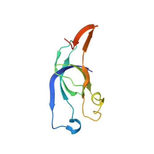
Two new high-resolution crystal structures of carboxysome pentamer proteins reveal high structural conservation of CcmL orthologs among distantly related cyanobacterial species.

Sutter, M.Wilson, S.C.Deutsch, S.Kerfeld, C.A.

(2013) Photosynth Res 118: 9-16

  • DOI: https://doi.org/10.1007/s11120-013-9909-z
  • Primary Citation of Related Structures:  
    4JVZ, 4JW0

  • PubMed Abstract: 

    Cyanobacteria have evolved a unique carbon fixation organelle known as the carboxysome that compartmentalizes the enzymes RuBisCO and carbonic anhydrase. This effectively increases the local CO2 concentration at the active site of RuBisCO and decreases its relatively unproductive side reaction with oxygen. Carboxysomes consist of a protein shell composed of hexameric and pentameric proteins arranged in icosahedral symmetry. Facets composed of hexameric proteins are connected at the vertices by pentameric proteins. Structurally homologous pentamers and hexamers are also found in heterotrophic bacteria where they form architecturally related microcompartments such as the Eut and Pdu organelles for the metabolism of ethanolamine and propanediol, respectively. Here we describe two new high-resolution structures of the pentameric shell protein CcmL from the cyanobacteria Thermosynechococcus elongatus and Gloeobacter violaceus and provide detailed analysis of their characteristics and comparison with related shell proteins.


  • Organizational Affiliation

    United States Department of Energy - Joint Genome Institute, Walnut Creek, CA, 94598, USA.


Macromolecules
Find similar proteins by:  (by identity cutoff)  |  3D Structure
Entity ID: 1
MoleculeChains Sequence LengthOrganismDetailsImage
Carbon dioxide concentrating mechanism proteinA [auth B],
B [auth A],
C,
D,
E
108Gloeobacter violaceus PCC 7421Mutation(s): 0 
Gene Names: ccmLgll2094
UniProt
Find proteins for Q7NIT8 (Gloeobacter violaceus (strain ATCC 29082 / PCC 7421))
Explore Q7NIT8 
Go to UniProtKB:  Q7NIT8
Entity Groups  
Sequence Clusters30% Identity50% Identity70% Identity90% Identity95% Identity100% Identity
UniProt GroupQ7NIT8
Sequence Annotations
Expand
  • Reference Sequence
Small Molecules
Ligands 1 Unique
IDChains Name / Formula / InChI Key2D Diagram3D Interactions
SO4
Query on SO4

Download Ideal Coordinates CCD File 
F [auth A]SULFATE ION
O4 S
QAOWNCQODCNURD-UHFFFAOYSA-L
Experimental Data & Validation

Experimental Data

  • Method: X-RAY DIFFRACTION
  • Resolution: 1.70 Å
  • R-Value Free: 0.191 
  • R-Value Work: 0.161 
  • R-Value Observed: 0.162 
  • Space Group: P 21 21 21
Unit Cell:
Length ( Å )Angle ( ˚ )
a = 74.377α = 90
b = 81.619β = 90
c = 89.349γ = 90
Software Package:
Software NamePurpose
ADSCdata collection
AutoSolphasing
PHENIXrefinement
XDSdata reduction
SCALAdata scaling

Structure Validation

View Full Validation Report


Entry History 

Deposition Data

Revision History  (Full details and data files)

  • Version 1.0: 2013-09-04
    Type: Initial release
  • Version 1.1: 2013-11-27
    Changes: Database references
  • Version 1.2: 2024-02-28
    Changes: Data collection, Database references, Derived calculations