Small molecule inhibition of the KRAS PDEd interaction impairs oncogenic KRAS signalling
Zimmermann, G., Papke, B., Ismail, S., Vartak, N., Chandra, A., Hoffmann, M., Hahn, S.A., Triola, G., Wittinghofer, A., Bastiaens, P.I., Waldmann, H.(2013) Nature 497: 638-642
- PubMed: 23698361 Search on PubMed
- DOI: https://doi.org/10.1038/nature12205
- Primary Citation Related Structures:
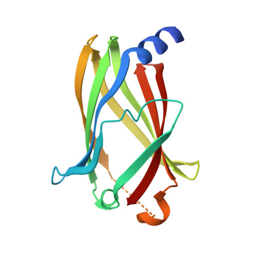
4JV6, 4JV8, 4JVB, 4JVF - PubMed Abstract:
The KRAS oncogene product is considered a major target in anticancer drug discovery. However, direct interference with KRAS signalling has not yet led to clinically useful drugs. Correct localization and signalling by farnesylated KRAS is regulated by the prenyl-binding protein PDEδ, which sustains the spatial organization of KRAS by facilitating its diffusion in the cytoplasm. Here we report that interfering with binding of mammalian PDEδ to KRAS by means of small molecules provides a novel opportunity to suppress oncogenic RAS signalling by altering its localization to endomembranes. Biochemical screening and subsequent structure-based hit optimization yielded inhibitors of the KRAS-PDEδ interaction that selectively bind to the prenyl-binding pocket of PDEδ with nanomolar affinity, inhibit oncogenic RAS signalling and suppress in vitro and in vivo proliferation of human pancreatic ductal adenocarcinoma cells that are dependent on oncogenic KRAS. Our findings may inspire novel drug discovery efforts aimed at the development of drugs targeting oncogenic RAS.
- Department of Chemical Biology, Max Planck Institute of Molecular Physiology, D-44227 Dortmund, Germany.
Organizational Affiliation: