4JVE | pdb_00004jve

Co-crystal structure of MDM2 with inhibitor (2R,3E)-2-[(2S,3R,6S)-2,3-bis(4-chlorophenyl)-6-(4-fluorobenzyl)-5-oxomorpholin-4-yl]pent-3-enoic acid


Experimental Data Snapshot

  • Method: X-RAY DIFFRACTION
  • Resolution: 2.30 Å
  • R-Value Free: 
    0.298 (Depositor), 0.336 (DCC) 
  • R-Value Work: 
    0.268 (Depositor), 0.268 (DCC) 

wwPDB Validation 3D Report Full Report

Validation slider image for 4JVE

Ligand Structure Quality Assessment 


This is version 1.2 of the entry. See complete history

Literature

Rational Design and Binding Mode Duality of MDM2-p53 Inhibitors.

Gonzalez-Lopez de Turiso, F.Sun, D.Rew, Y.Bartberger, M.D.Beck, H.P.Canon, J.Chen, A.Chow, D.Correll, T.L.Huang, X.Julian, L.D.Kayser, F.Lo, M.C.Long, A.M.McMinn, D.Oliner, J.D.Osgood, T.Powers, J.P.Saiki, A.Y.Schneider, S.Shaffer, P.Xiao, S.H.Yakowec, P.Yan, X.Ye, Q.Yu, D.Zhao, X.Zhou, J.Medina, J.C.Olson, S.H.

(2013) J Med Chem 56: 4053-4070

  • DOI: https://doi.org/10.1021/jm400293z
  • Primary Citation Related Structures: 
    4JV7, 4JV9, 4JVE, 4JVR, 4JWR

  • PubMed Abstract: 

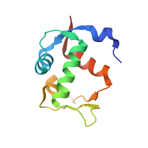
    Structural analysis of both the MDM2-p53 protein-protein interaction and several small molecules bound to MDM2 led to the design and synthesis of tetrasubstituted morpholinone 10, an MDM2 inhibitor with a biochemical IC50 of 1.0 μM. The cocrystal structure of 10 with MDM2 inspired two independent optimization strategies and resulted in the discovery of morpholinones 16 and 27 possessing distinct binding modes. Both analogues were potent MDM2 inhibitors in biochemical and cellular assays, and morpholinone 27 (IC50 = 0.10 μM) also displayed suitable PK profile for in vivo animal experiments. A pharmacodynamic (PD) experiment in mice implanted with human SJSA-1 tumors showed p21(WAF1) mRNA induction (2.7-fold over vehicle) upon oral dosing of 27 at 300 mg/kg.


  • Organizational Affiliation
    • Department of Therapeutic Discovery, Amgen Inc. , 1120 Veterans Boulevard, South San Francisco, California 94080, USA.

Macromolecule Content 

  • Total Structure Weight: 11.68 kDa 
  • Atom Count: 830 
  • Modeled Residue Count: 92 
  • Deposited Residue Count: 96 
  • Unique protein chains: 1

Macromolecules

Find similar proteins by:|  3D Structure
Entity ID: 1
MoleculeChains  Sequence LengthOrganismDetailsImage
E3 ubiquitin-protein ligase Mdm296Homo sapiensMutation(s): 0 
Gene Names: MDM2
EC: 6.3.2 (PDB Primary Data), 2.3.2.27 (UniProt)
UniProt & NIH Common Fund Data Resources
Find proteins for Q00987 (Homo sapiens)
Explore Q00987 
Go to UniProtKB:  Q00987
PHAROS:  Q00987
GTEx:  ENSG00000135679 
Entity Groups
Sequence Clusters30% Identity50% Identity70% Identity90% Identity95% Identity100% Identity
UniProt GroupQ00987
Sequence Annotations
Expand
Reference Sequence

Small Molecules

Ligands 1 Unique
IDChains Name / Formula / InChI Key2D Diagram3D Interactions
1MQ

Query on 1MQ


Download:Ideal Coordinates CCD File
B [auth A](2R,3E)-2-[(2S,3R,6S)-2,3-bis(4-chlorophenyl)-6-(4-fluorobenzyl)-5-oxomorpholin-4-yl]pent-3-enoic acid
C28 H24 Cl2 F N O4
KFJHASQDDHBQDA-GYEZOCHYSA-N

Experimental Data & Validation

Experimental Data

  • Method: X-RAY DIFFRACTION
  • Resolution: 2.30 Å
  • R-Value Free:  0.298 (Depositor), 0.336 (DCC) 
  • R-Value Work:  0.268 (Depositor), 0.268 (DCC) 
Space Group: P 41 21 2
Unit Cell:
Length ( Å )Angle ( ˚ )
a = 42.014α = 90
b = 42.014β = 90
c = 119.418γ = 90
Software Package:
Software NamePurpose
HKL-2000data collection
AMoREphasing
CNSrefinement
DENZOdata reduction
SCALEPACKdata scaling

Structure Validation

View Full Validation Report


Ligand Structure Quality Assessment 


Entry History 

Revision History  (Full details and data files)

  • Version 1.0: 2013-05-01
    Type: Initial release
  • Version 1.1: 2013-06-05
    Changes: Database references
  • Version 1.2: 2024-11-06
    Changes: Data collection, Database references, Derived calculations, Structure summary