4JQU | pdb_00004jqu

Crystal structure of Ubc7p in complex with the U7BR of Cue1p


Experimental Data Snapshot

  • Method: X-RAY DIFFRACTION
  • Resolution: 1.81 Å
  • R-Value Free: 
    0.225 (Depositor), 0.228 (DCC) 
  • R-Value Work: 
    0.184 (Depositor), 0.183 (DCC) 
  • R-Value Observed: 
    0.186 (Depositor) 

Starting Model: experimental
View more details

wwPDB Validation 3D Report Full Report

Validation slider image for 4JQU

This is version 1.4 of the entry. See complete history

Literature

A Structurally Unique E2-Binding Domain Activates Ubiquitination by the ERAD E2, Ubc7p, through Multiple Mechanisms.

Metzger, M.B.Liang, Y.H.Das, R.Mariano, J.Li, S.Li, J.Kostova, Z.Byrd, R.A.Ji, X.Weissman, A.M.

(2013) Mol Cell 50: 516-527

  • DOI: https://doi.org/10.1016/j.molcel.2013.04.004
  • Primary Citation Related Structures: 
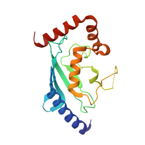
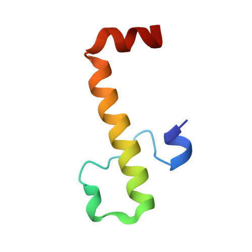
    4JQU

  • PubMed Abstract: 

    Cue1p is an integral component of yeast endoplasmic reticulum (ER)-associated degradation (ERAD) ubiquitin ligase (E3) complexes. It tethers the ERAD ubiquitin-conjugating enzyme (E2), Ubc7p, to the ER and prevents its degradation, and also activates Ubc7p via unknown mechanisms. We have now determined the crystal structure of the Ubc7p-binding region (U7BR) of Cue1p with Ubc7p. The U7BR is a unique E2-binding domain that includes three α-helices that interact extensively with the "backside" of Ubc7p. Residues essential for E2 binding are also required for activation of Ubc7p and for ERAD. We establish that the U7BR stimulates both RING-independent and RING-dependent ubiquitin transfer from Ubc7p. Moreover, the U7BR enhances ubiquitin-activating enzyme (E1)-mediated charging of Ubc7p with ubiquitin. This demonstrates that an essential component of E3 complexes can simultaneously bind to E2 and enhance its loading with ubiquitin. These findings provide mechanistic insights into how ubiquitination can be stimulated.


  • Organizational Affiliation
    • Laboratory of Protein Dynamics and Signaling, Center for Cancer Research, National Cancer Institute, Frederick, MD 21702-1201, USA.

Macromolecule Content 

  • Total Structure Weight: 25.67 kDa 
  • Atom Count: 1,993 
  • Modeled Residue Count: 217 
  • Deposited Residue Count: 223 
  • Unique protein chains: 2

Macromolecules

Find similar proteins by:|  3D Structure
Entity ID: 1
MoleculeChains  Sequence LengthOrganismDetailsImage
Ubiquitin-conjugating enzyme E2 7169Saccharomyces cerevisiae S288CMutation(s): 0 
Gene Names: QRI8UBC7Ubc7pYM9711.12YMR022W
EC: 6.3.2.19 (PDB Primary Data), 2.3.2.23 (UniProt)
UniProt
Find proteins for Q02159 (Saccharomyces cerevisiae (strain ATCC 204508 / S288c))
Explore Q02159 
Go to UniProtKB:  Q02159
Entity Groups
Sequence Clusters30% Identity50% Identity70% Identity90% Identity95% Identity100% Identity
UniProt GroupQ02159
Sequence Annotations
Expand
Reference Sequence
Find similar proteins by:|  3D Structure
Entity ID: 2
MoleculeChains  Sequence LengthOrganismDetailsImage
Coupling of ubiquitin conjugation to ER degradation protein 154Saccharomyces cerevisiae S288CMutation(s): 0 
Gene Names: CUE1Cue1pKIS4YM8156.06YMR264W
UniProt
Find proteins for P38428 (Saccharomyces cerevisiae (strain ATCC 204508 / S288c))
Explore P38428 
Go to UniProtKB:  P38428
Entity Groups
Sequence Clusters30% Identity50% Identity70% Identity90% Identity95% Identity100% Identity
UniProt GroupP38428
Sequence Annotations
Expand
Reference Sequence

Experimental Data & Validation

Experimental Data

  • Method: X-RAY DIFFRACTION
  • Resolution: 1.81 Å
  • R-Value Free:  0.225 (Depositor), 0.228 (DCC) 
  • R-Value Work:  0.184 (Depositor), 0.183 (DCC) 
  • R-Value Observed: 0.186 (Depositor) 
Space Group: P 21 21 21
Unit Cell:
Length ( Å )Angle ( ˚ )
a = 46.81α = 90
b = 48.463β = 90
c = 95.734γ = 90
Software Package:
Software NamePurpose
DENZOdata reduction
SCALEPACKdata scaling
PHASERphasing
PHENIXrefinement
PDB_EXTRACTdata extraction
HKL-3000data reduction
HKL-3000data scaling

Structure Validation

View Full Validation Report


Entry History 

Deposition Data

Revision History  (Full details and data files)

  • Version 1.0: 2013-05-29
    Type: Initial release
  • Version 1.1: 2013-06-19
    Changes: Database references
  • Version 1.2: 2017-11-15
    Changes: Refinement description
  • Version 1.3: 2023-08-30
    Changes: Data collection, Database references, Derived calculations, Structure summary
  • Version 1.4: 2023-09-20
    Changes: Refinement description