The first crystal structure of NAD-dependent 3-dehydro-2-deoxy-D-gluconate dehydrogenase from Thermus thermophilus HB8
Pampa, K.J., Lokanath, N.K., Kunishima, N., Ravishnkar Rai, V.(2014) Acta Crystallogr D Biol Crystallogr 70: 994-1004
- PubMed: 24699644 Search on PubMed
- DOI: https://doi.org/10.1107/S1399004713034925
- Primary Citation Related Structures:
2EKP, 2EKQ, 4JP2, 4JP3 - PubMed Abstract:
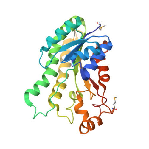
2-Keto-3-deoxygluconate (KDG) is one of the important intermediates in pectin metabolism. An enzyme involved in this pathway, 3-dehydro-3-deoxy-D-gluconate 5-dehydrogenase (DDGDH), has been identified which converts 2,5-diketo-3-deoxygluconate to KDG. The enzyme is a member of the short-chain dehydrogenase (SDR) family. To gain insight into the function of this enzyme at the molecular level, the first crystal structure of DDGDH from Thermus thermophilus HB8 has been determined in the apo form, as well as in complexes with the cofactor and with citrate, by X-ray diffraction methods. The crystal structures reveal a tight tetrameric oligomerization. The secondary-structural elements and catalytically important residues of the enzyme were highly conserved amongst the proteins of the NAD(P)-dependent SDR family. The DDGDH protomer contains a dinucleotide-binding fold which binds the coenzyme NAD(+) in an intersubunit cleft; hence, the observed oligomeric state might be important for the catalytic function. This enzyme prefers NAD(H) rather than NADP(H) as the physiological cofactor. A structural comparison of DDGDH with mouse lung carbonyl reductase suggests that a significant difference in the α-loop-α region of this enzyme is associated with the coenzyme specificity. The structural data allow a detailed understanding of the functional role of the conserved catalytic triad (Ser129-Tyr144-Lys148) in cofactor and substrate recognition, thus providing substantial insights into DDGDH catalysis. From analysis of the three-dimensional structure, intersubunit hydrophobic interactions were found to be important for enzyme oligomerization and thermostability.
- Department of Studies in Microbiology, University of Mysore, Manasagangotri, Mysore 570 006, India.
Organizational Affiliation: