4JMZ | pdb_00004jmz

Crystal structure of Cytochrome C Peroxidase W191G-Gateless in complex with N-methyl-1H-benzimidazol-2-amine


Experimental Data Snapshot

  • Method: X-RAY DIFFRACTION
  • Resolution: 1.82 Å
  • R-Value Free: 
    0.182 (Depositor), 0.193 (DCC) 
  • R-Value Work: 
    0.145 (Depositor), 0.157 (DCC) 
  • R-Value Observed: 
    0.147 (Depositor) 

wwPDB Validation 3D Report Full Report

Validation slider image for 4JMZ

Ligand Structure Quality Assessment 


This is version 1.2 of the entry. See complete history

Literature

Roles for ordered and bulk solvent in ligand recognition and docking in two related cavities.

Barelier, S.Boyce, S.E.Fish, I.Fischer, M.Goodin, D.B.Shoichet, B.K.

(2013) PLoS One 8: e69153-e69153

  • DOI: https://doi.org/10.1371/journal.pone.0069153
  • Primary Citation Related Structures: 
    4JMZ

  • PubMed Abstract: 

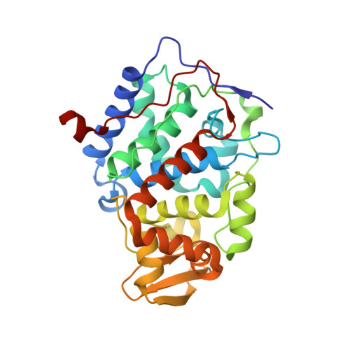
    A key challenge in structure-based discovery is accounting for modulation of protein-ligand interactions by ordered and bulk solvent. To investigate this, we compared ligand binding to a buried cavity in Cytochrome c Peroxidase (CcP), where affinity is dominated by a single ionic interaction, versus a cavity variant partly opened to solvent by loop deletion. This opening had unexpected effects on ligand orientation, affinity, and ordered water structure. Some ligands lost over ten-fold in affinity and reoriented in the cavity, while others retained their geometries, formed new interactions with water networks, and improved affinity. To test our ability to discover new ligands against this opened site prospectively, a 534,000 fragment library was docked against the open cavity using two models of ligand solvation. Using an older solvation model that prioritized many neutral molecules, three such uncharged docking hits were tested, none of which was observed to bind; these molecules were not highly ranked by the new, context-dependent solvation score. Using this new method, another 15 highly-ranked molecules were tested for binding. In contrast to the previous result, 14 of these bound detectably, with affinities ranging from 8 µM to 2 mM. In crystal structures, four of these new ligands superposed well with the docking predictions but two did not, reflecting unanticipated interactions with newly ordered waters molecules. Comparing recognition between this open cavity and its buried analog begins to isolate the roles of ordered solvent in a system that lends itself readily to prospective testing and that may be broadly useful to the community.


  • Organizational Affiliation
    • Department of Pharmaceutical Chemistry, University of California San Francisco, San Francisco, California, USA.

Macromolecule Content 

  • Total Structure Weight: 33.69 kDa 
  • Atom Count: 2,956 
  • Modeled Residue Count: 289 
  • Deposited Residue Count: 289 
  • Unique protein chains: 1

Macromolecules

Find similar proteins by:|  3D Structure
Entity ID: 1
MoleculeChains  Sequence LengthOrganismDetailsImage
Cytochrome c peroxidase289Saccharomyces cerevisiae RM11-1aMutation(s): 2 
Gene Names: CCP1 CCP CPO YKR066CSCRG_04081
EC: 1.11.1.5 (PDB Primary Data), 1.11.1 (UniProt)
UniProt
Find proteins for B3LRE1 (Saccharomyces cerevisiae (strain RM11-1a))
Explore B3LRE1 
Go to UniProtKB:  B3LRE1
Entity Groups
Sequence Clusters30% Identity50% Identity70% Identity90% Identity95% Identity100% Identity
UniProt GroupB3LRE1
Sequence Annotations
Expand
Reference Sequence

Small Molecules

Ligands 2 Unique
IDChains Name / Formula / InChI Key2D Diagram3D Interactions
HEM

Query on HEM


Download:Ideal Coordinates CCD File
B [auth A]PROTOPORPHYRIN IX CONTAINING FE
C34 H32 Fe N4 O4
KABFMIBPWCXCRK-RGGAHWMASA-L
1M2

Query on 1M2


Download:Ideal Coordinates CCD File
C [auth A]N-methyl-1H-benzimidazol-2-amine
C8 H9 N3
ALBBQFWHUCTZFM-UHFFFAOYSA-N

Experimental Data & Validation

Experimental Data

  • Method: X-RAY DIFFRACTION
  • Resolution: 1.82 Å
  • R-Value Free:  0.182 (Depositor), 0.193 (DCC) 
  • R-Value Work:  0.145 (Depositor), 0.157 (DCC) 
  • R-Value Observed: 0.147 (Depositor) 
Space Group: P 21 21 21
Unit Cell:
Length ( Å )Angle ( ˚ )
a = 105.15α = 90
b = 74.57β = 90
c = 51.18γ = 90
Software Package:
Software NamePurpose
REFMACrefinement
PDB_EXTRACTdata extraction
ADSCdata collection
XDSdata reduction
XDSdata scaling

Structure Validation

View Full Validation Report


Ligand Structure Quality Assessment 


Entry History 

Deposition Data

Revision History  (Full details and data files)

  • Version 1.0: 2013-05-01
    Type: Initial release
  • Version 1.1: 2013-08-28
    Changes: Database references
  • Version 1.2: 2024-02-28
    Changes: Data collection, Database references, Derived calculations