4JLG | pdb_00004jlg

SETD7 in complex with inhibitor (R)-PFI-2 and S-adenosyl-methionine


Experimental Data Snapshot

  • Method: X-RAY DIFFRACTION
  • Resolution: 1.90 Å
  • R-Value Free: 
    0.237 (Depositor), 0.236 (DCC) 
  • R-Value Work: 
    0.211 (Depositor), 0.208 (DCC) 
  • R-Value Observed: 
    0.212 (Depositor) 

Starting Model: experimental
View more details

wwPDB Validation 3D Report Full Report

Validation slider image for 4JLG

Ligand Structure Quality Assessment 


This is version 1.3 of the entry. See complete history

Literature

(R)-PFI-2 is a potent and selective inhibitor of SETD7 methyltransferase activity in cells.

Barsyte-Lovejoy, D.Li, F.Oudhoff, M.J.Tatlock, J.H.Dong, A.Zeng, H.Wu, H.Freeman, S.A.Schapira, M.Senisterra, G.A.Kuznetsova, E.Marcellus, R.Allali-Hassani, A.Kennedy, S.Lambert, J.P.Couzens, A.L.Aman, A.Gingras, A.C.Al-Awar, R.Fish, P.V.Gerstenberger, B.S.Roberts, L.Benn, C.L.Grimley, R.L.Braam, M.J.Rossi, F.M.Sudol, M.Brown, P.J.Bunnage, M.E.Owen, D.R.Zaph, C.Vedadi, M.Arrowsmith, C.H.

(2014) Proc Natl Acad Sci U S A 111: 12853-12858

  • DOI: https://doi.org/10.1073/pnas.1407358111
  • Primary Citation Related Structures: 
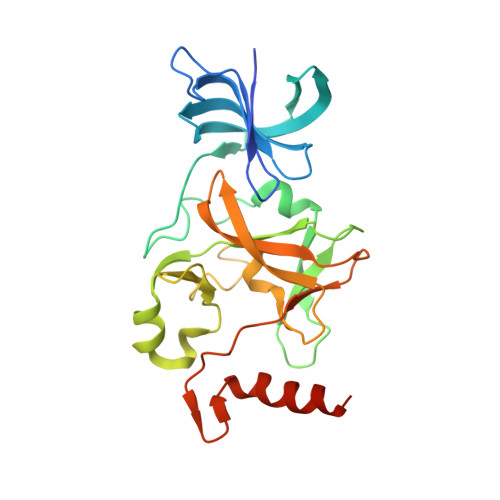
    4JLG

  • PubMed Abstract: 

    SET domain containing (lysine methyltransferase) 7 (SETD7) is implicated in multiple signaling and disease related pathways with a broad diversity of reported substrates. Here, we report the discovery of (R)-PFI-2-a first-in-class, potent (Ki (app) = 0.33 nM), selective, and cell-active inhibitor of the methyltransferase activity of human SETD7-and its 500-fold less active enantiomer, (S)-PFI-2. (R)-PFI-2 exhibits an unusual cofactor-dependent and substrate-competitive inhibitory mechanism by occupying the substrate peptide binding groove of SETD7, including the catalytic lysine-binding channel, and by making direct contact with the donor methyl group of the cofactor, S-adenosylmethionine. Chemoproteomics experiments using a biotinylated derivative of (R)-PFI-2 demonstrated dose-dependent competition for binding to endogenous SETD7 in MCF7 cells pretreated with (R)-PFI-2. In murine embryonic fibroblasts, (R)-PFI-2 treatment phenocopied the effects of Setd7 deficiency on Hippo pathway signaling, via modulation of the transcriptional coactivator Yes-associated protein (YAP) and regulation of YAP target genes. In confluent MCF7 cells, (R)-PFI-2 rapidly altered YAP localization, suggesting continuous and dynamic regulation of YAP by the methyltransferase activity of SETD7. These data establish (R)-PFI-2 and related compounds as a valuable tool-kit for the study of the diverse roles of SETD7 in cells and further validate protein methyltransferases as a druggable target class.


  • Organizational Affiliation
    • Structural Genomics Consortium, University of Toronto, Toronto, ON, Canada M5G 1L7; carrow@uhnres.utoronto.ca m.vedadi@utoronto.ca d.barsyte@utoronto.ca.

Macromolecule Content 

  • Total Structure Weight: 61.16 kDa 
  • Atom Count: 4,374 
  • Modeled Residue Count: 493 
  • Deposited Residue Count: 528 
  • Unique protein chains: 1

Macromolecules

Find similar proteins by:|  3D Structure
Entity ID: 1
MoleculeChains  Sequence LengthOrganismDetailsImage
Histone-lysine N-methyltransferase SETD7
A, B
264Homo sapiensMutation(s): 0 
Gene Names: SETD7KIAA1717KMT7SET7SET9
EC: 2.1.1.43 (PDB Primary Data), 2.1.1.364 (UniProt)
UniProt & NIH Common Fund Data Resources
Find proteins for Q8WTS6 (Homo sapiens)
Explore Q8WTS6 
Go to UniProtKB:  Q8WTS6
PHAROS:  Q8WTS6
GTEx:  ENSG00000145391 
Entity Groups
Sequence Clusters30% Identity50% Identity70% Identity90% Identity95% Identity100% Identity
UniProt GroupQ8WTS6
Sequence Annotations
Expand
Reference Sequence

Small Molecules

Ligands 3 Unique
IDChains Name / Formula / InChI Key2D Diagram3D Interactions
1L8

Query on 1L8


Download:Ideal Coordinates CCD File
D [auth A],
W [auth B]
8-fluoro-N-{(2R)-1-oxo-1-(pyrrolidin-1-yl)-3-[3-(trifluoromethyl)phenyl]propan-2-yl}-1,2,3,4-tetrahydroisoquinoline-6-sulfonamide
C23 H25 F4 N3 O3 S
JCKGSPAAPQRPBW-OAQYLSRUSA-N
SAM

Query on SAM


Download:Ideal Coordinates CCD File
C [auth A],
V [auth B]
S-ADENOSYLMETHIONINE
C15 H22 N6 O5 S
MEFKEPWMEQBLKI-FCKMPRQPSA-N
UNX

Query on UNX


Download:Ideal Coordinates CCD File
AA [auth B]
BA [auth B]
CA [auth B]
DA [auth B]
E [auth A]
AA [auth B],
BA [auth B],
CA [auth B],
DA [auth B],
E [auth A],
EA [auth B],
F [auth A],
G [auth A],
H [auth A],
I [auth A],
J [auth A],
K [auth A],
L [auth A],
M [auth A],
N [auth A],
O [auth A],
P [auth A],
Q [auth A],
R [auth A],
S [auth A],
T [auth A],
U [auth A],
X [auth B],
Y [auth B],
Z [auth B]
UNKNOWN ATOM OR ION
X
Binding Affinity Annotations 
IDSourceBinding Affinity
1L8 BindingDB:  4JLG Ki: 0.33 (nM) from 1 assay(s)
Kd: 4.2 (nM) from 1 assay(s)
IC50: 2 (nM) from 1 assay(s)

Experimental Data & Validation

Experimental Data

  • Method: X-RAY DIFFRACTION
  • Resolution: 1.90 Å
  • R-Value Free:  0.237 (Depositor), 0.236 (DCC) 
  • R-Value Work:  0.211 (Depositor), 0.208 (DCC) 
  • R-Value Observed: 0.212 (Depositor) 
Space Group: P 1
Unit Cell:
Length ( Å )Angle ( ˚ )
a = 46.742α = 83.33
b = 57.984β = 71.42
c = 64.932γ = 70.93
Software Package:
Software NamePurpose
SCALEPACKdata scaling
REFMACrefinement
PDB_EXTRACTdata extraction
JBluIce-EPICSdata collection
HKL-2000data reduction
PHASESphasing

Structure Validation

View Full Validation Report


Ligand Structure Quality Assessment 


Entry History 

Revision History  (Full details and data files)

  • Version 1.0: 2013-04-17
    Type: Initial release
  • Version 1.1: 2014-04-09
    Changes: Structure summary
  • Version 1.2: 2015-02-18
    Changes: Database references
  • Version 1.3: 2023-09-20
    Changes: Data collection, Database references, Derived calculations, Refinement description