4JHZ | pdb_00004jhz

Structure of E. coli beta-Glucuronidase bound with a novel, potent inhibitor 2-[4-(1,3-benzodioxol-5-ylmethyl)piperazin-1-yl]-N-[(1S,2S,5S)-2,5-dimethoxycyclohexyl]acetamide


Experimental Data Snapshot

  • Method: X-RAY DIFFRACTION
  • Resolution: 2.83 Å
  • R-Value Free: 
    0.247 (Depositor), 0.244 (DCC) 
  • R-Value Work: 
    0.196 (Depositor), 0.200 (DCC) 
  • R-Value Observed: 
    0.198 (Depositor) 

Starting Model: experimental
View more details

wwPDB Validation 3D Report Full Report

Validation slider image for 4JHZ

Ligand Structure Quality Assessment 


This is version 1.3 of the entry. See complete history

Literature

Molecular Insights into Microbial beta-Glucuronidase Inhibition to Abrogate CPT-11 Toxicity.

Roberts, A.B.Wallace, B.D.Venkatesh, M.K.Mani, S.Redinbo, M.R.

(2013) Mol Pharmacol 84: 208-217

  • DOI: https://doi.org/10.1124/mol.113.085852
  • Primary Citation Related Structures: 
    4JHZ

  • PubMed Abstract: 

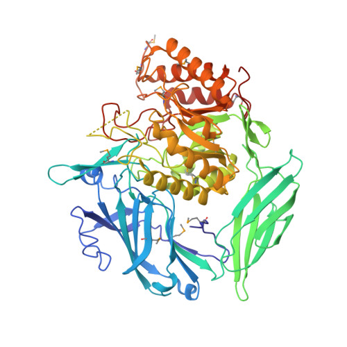
    Bacterial β-glucuronidases expressed by the symbiotic intestinal microbiota appear to play important roles in drug-induced epithelial cell toxicity in the gastrointestinal (GI) tract. For the anticancer drug CPT-11 (irinotecan) and the nonsteroidal anti-inflammatory drug diclofenac, it has been shown that removal of the glucuronide moieties from drug metabolites by bacterial β-glucuronidases in the GI lumen can significantly damage the intestinal epithelium. Furthermore, selective disruption of bacterial β-glucuronidases by small molecule inhibitors alleviates these side effects, which, for CPT-11 {7-ethyl-10-[4-(1-piperidino)-1-piperidino]}, can be dose limiting. Here we characterize novel microbial β-glucuronidase inhibitors that inhibit Escherichia coli β-glucuronidase in vitro with Ki values between 180 nM and 2 μM, and disrupt the enzyme in E. coli cells, with EC50 values as low as 300 nM. All compounds are selective for E. coli β-glucuronidase without inhibiting purified mammalian β-glucuronidase, and they do not impact the survival of either bacterial or mammalian cells. The 2.8 Å resolution crystal structure of one inhibitor bound to E. coli β-glucuronidase demonstrates that it contacts and orders only a portion of the "bacterial loop" present in microbial, but not mammalian, β-glucuronidases. The most potent compound examined in this group was found to protect mice against CPT-11-induced diarrhea. Taken together, these data advance our understanding of the chemical and structural basis of selective microbial β-glucuronidase inhibition, which may improve human drug efficacy and toxicity.


  • Organizational Affiliation
    • Departments of Biochemistry, Chemistry and Microbiology, University of North Carolina at Chapel Hill, NC, USA.

Macromolecule Content 

  • Total Structure Weight: 139.04 kDa 
  • Atom Count: 9,613 
  • Modeled Residue Count: 1,192 
  • Deposited Residue Count: 1,206 
  • Unique protein chains: 1

Macromolecules

Find similar proteins by:|  3D Structure
Entity ID: 1
MoleculeChains  Sequence LengthOrganismDetailsImage
Beta-glucuronidase
A, B
603Escherichia coli K-12Mutation(s): 0 
Gene Names: b1617gurAgusAJW1609uidA
EC: 3.2.1.31
UniProt
Find proteins for P05804 (Escherichia coli (strain K12))
Explore P05804 
Go to UniProtKB:  P05804
Entity Groups
Sequence Clusters30% Identity50% Identity70% Identity90% Identity95% Identity100% Identity
UniProt GroupP05804
Sequence Annotations
Expand
Reference Sequence

Small Molecules

Ligands 1 Unique
IDChains Name / Formula / InChI Key2D Diagram3D Interactions
1KV

Query on 1KV


Download:Ideal Coordinates CCD File
C [auth A],
D [auth B]
2-[4-(1,3-benzodioxol-5-ylmethyl)piperazin-1-yl]-N-[(1S,2S,5S)-2,5-dimethoxycyclohexyl]acetamide
C22 H33 N3 O5
AOKPSAUXHJHABU-QYZOEREBSA-N
Modified Residues  1 Unique
IDChains TypeFormula2D DiagramParent
MSE
Query on MSE
A, B
L-PEPTIDE LINKINGC5 H11 N O2 SeMET

Experimental Data & Validation

Experimental Data

  • Method: X-RAY DIFFRACTION
  • Resolution: 2.83 Å
  • R-Value Free:  0.247 (Depositor), 0.244 (DCC) 
  • R-Value Work:  0.196 (Depositor), 0.200 (DCC) 
  • R-Value Observed: 0.198 (Depositor) 
Space Group: C 1 2 1
Unit Cell:
Length ( Å )Angle ( ˚ )
a = 168.305α = 90
b = 77.408β = 124.84
c = 126.291γ = 90
Software Package:
Software NamePurpose
HKL-2000data collection
PHASERphasing
PHENIXrefinement
HKL-2000data reduction
HKL-2000data scaling

Structure Validation

View Full Validation Report


Ligand Structure Quality Assessment 


Entry History 

Deposition Data

Revision History  (Full details and data files)

  • Version 1.0: 2013-08-28
    Type: Initial release
  • Version 1.1: 2023-09-20
    Changes: Data collection, Database references, Derived calculations, Refinement description
  • Version 1.2: 2023-12-06
    Changes: Data collection
  • Version 1.3: 2024-11-20
    Changes: Structure summary