4JHR | pdb_00004jhr

An auto-inhibited conformation of LGN reveals a distinct interaction mode between GoLoco motifs and TPR motifs


Experimental Data Snapshot

  • Method: X-RAY DIFFRACTION
  • Resolution: 2.80 Å
  • R-Value Free: 
    0.311 (Depositor), 0.327 (DCC) 
  • R-Value Work: 
    0.250 (Depositor), 0.266 (DCC) 
  • R-Value Observed: 
    0.253 (Depositor) 

Starting Model: experimental
View more details

wwPDB Validation 3D Report Full Report

Validation slider image for 4JHR

This is version 1.4 of the entry. See complete history

Literature

An autoinhibited conformation of LGN reveals a distinct interaction mode between GoLoco motifs and TPR motifs

Pan, Z.Zhu, J.Shang, Y.Wei, Z.Jia, M.Xia, C.Wen, W.Wang, W.Zhang, M.

(2013) Structure 21: 1007-1017

  • DOI: https://doi.org/10.1016/j.str.2013.04.005
  • Primary Citation Related Structures: 
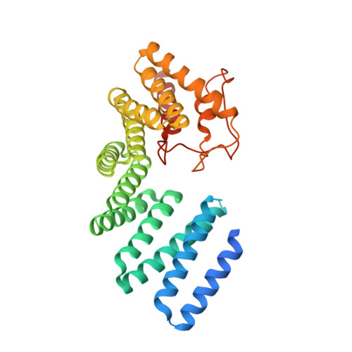
    4JHR

  • PubMed Abstract: 

    LGN plays essential roles in asymmetric cell divisions via its N-terminal TPR-motif-mediated binding to mInsc and NuMA. This scaffolding activity requires the release of the autoinhibited conformation of LGN by binding of Gα(i) to its C-terminal GoLoco (GL) motifs. The interaction between the GL and TPR motifs of LGN represents a distinct GL/target binding mode with an unknown mechanism. Here, we show that two consecutive GL motifs of LGN form a minimal TPR-motif-binding unit. GL12 and GL34 bind to TPR0-3 and TPR4-7, respectively. The crystal structure of a truncated LGN reveals that GL34 forms a pair of parallel α helices and binds to the concave surface of TPR4-7, thereby preventing LGN from binding to other targets. Importantly, the GLs bind to TPR motifs with a mode distinct from that observed in the GL/Gα(i)·GDP complexes. Our results also indicate that multiple and orphan GL motif proteins likely respond to G proteins with distinct mechanisms.


  • Organizational Affiliation
    • Shanghai Key Laboratory of Molecular Catalysis and Innovative Materials, Department of Chemistry and Institutes of Biomedical Sciences, Fudan University, Shanghai, China.

Macromolecule Content 

  • Total Structure Weight: 73.76 kDa 
  • Atom Count: 4,249 
  • Modeled Residue Count: 579 
  • Deposited Residue Count: 666 
  • Unique protein chains: 1

Macromolecules

Find similar proteins by:|  3D Structure
Entity ID: 1
MoleculeChains  Sequence LengthOrganismDetailsImage
G-protein-signaling modulator 2
A, B
333Mus musculusMutation(s): 0 
Gene Names: Gpsm2LgnPins
UniProt & NIH Common Fund Data Resources
Find proteins for Q8VDU0 (Mus musculus)
Explore Q8VDU0 
Go to UniProtKB:  Q8VDU0
IMPC:  MGI:1923373
Entity Groups
Sequence Clusters30% Identity50% Identity70% Identity90% Identity95% Identity100% Identity
UniProt GroupQ8VDU0
Sequence Annotations
Expand
Reference Sequence

Experimental Data & Validation

Experimental Data

  • Method: X-RAY DIFFRACTION
  • Resolution: 2.80 Å
  • R-Value Free:  0.311 (Depositor), 0.327 (DCC) 
  • R-Value Work:  0.250 (Depositor), 0.266 (DCC) 
  • R-Value Observed: 0.253 (Depositor) 
Space Group: P 2 21 21
Unit Cell:
Length ( Å )Angle ( ˚ )
a = 72.213α = 90
b = 81.803β = 90
c = 106.942γ = 90
Software Package:
Software NamePurpose
HKL-2000data collection
PHASESphasing
PHENIXrefinement
HKL-2000data reduction
HKL-2000data scaling

Structure Validation

View Full Validation Report


Entry History 

Deposition Data

Revision History  (Full details and data files)

  • Version 1.0: 2013-06-05
    Type: Initial release
  • Version 1.1: 2013-09-04
    Changes: Database references
  • Version 1.2: 2017-08-02
    Changes: Refinement description, Source and taxonomy
  • Version 1.3: 2023-11-08
    Changes: Data collection, Database references, Refinement description
  • Version 1.4: 2024-10-30
    Changes: Structure summary