4JHQ | pdb_00004jhq

Crystal structure of avidin - 6-(6-biotinamidohexanamido)hexanoylferrocene complex


Experimental Data Snapshot

  • Method: X-RAY DIFFRACTION
  • Resolution: 1.99 Å
  • R-Value Free: 
    0.210 (Depositor), 0.209 (DCC) 
  • R-Value Work: 
    0.162 (Depositor), 0.161 (DCC) 
  • R-Value Observed: 
    0.165 (Depositor) 

Starting Model: experimental
View more details

wwPDB Validation 3D Report Full Report

Validation slider image for 4JHQ

Ligand Structure Quality Assessment 


This is version 1.3 of the entry. See complete history

Literature

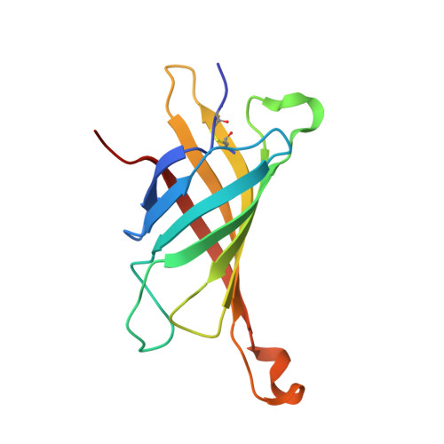
Ferrocene-Biotin Conjugates Targeting Cancer Cells: Synthesis, Interaction with Avidin, Cytotoxic Properties and the Crystal Structure of the Complex of Avidin with a Biotin-Linker-Ferrocene Conjugate

Plazuk, D.Zakrzewski, J.Salmain, M.Blauz, A.Rychlik, B.Strzelczyk, P.Bujacz, A.Bujacz, G.

(2013) Organometallics 32: 5774-5783

Macromolecule Content 

  • Total Structure Weight: 30.35 kDa 
  • Atom Count: 2,175 
  • Modeled Residue Count: 247 
  • Deposited Residue Count: 256 
  • Unique protein chains: 1

Macromolecules

Find similar proteins by:|  3D Structure
Entity ID: 1
MoleculeChains  Sequence LengthOrganismDetailsImage
Avidin
A, B
128Gallus gallusMutation(s): 0 
UniProt
Find proteins for P02701 (Gallus gallus)
Explore P02701 
Go to UniProtKB:  P02701
Entity Groups
Sequence Clusters30% Identity50% Identity70% Identity90% Identity95% Identity100% Identity
UniProt GroupP02701
Glycosylation
Glycosylation Sites: 1
Sequence Annotations
Expand
Reference Sequence

Small Molecules

Ligands 2 Unique
IDChains Name / Formula / InChI Key2D Diagram3D Interactions
B6F

Query on B6F


Download:Ideal Coordinates CCD File
C [auth A],
E [auth B]
[(1,2,3,4,5-eta)-cyclopentadienyl][(1,2,3,4,5-eta)-{6-[(6-{[5-(2-oxohexahydro-1H-thieno[3,4-d]imidazol-4-yl)pentanoyl]amino}hexanoyl)amino]hexanoyl}cyclopentadienyl]iron
C32 H38 Fe N4 O4 S
GEJZIMWXLQTIFK-WOZYLDAHSA-N
NAG

Query on NAG


Download:Ideal Coordinates CCD File
D [auth A],
F [auth B]
2-acetamido-2-deoxy-beta-D-glucopyranose
C8 H15 N O6
OVRNDRQMDRJTHS-FMDGEEDCSA-N

Experimental Data & Validation

Experimental Data

  • Method: X-RAY DIFFRACTION
  • Resolution: 1.99 Å
  • R-Value Free:  0.210 (Depositor), 0.209 (DCC) 
  • R-Value Work:  0.162 (Depositor), 0.161 (DCC) 
  • R-Value Observed: 0.165 (Depositor) 
Space Group: P 21 21 2
Unit Cell:
Length ( Å )Angle ( ˚ )
a = 69.693α = 90
b = 78.656β = 90
c = 42.762γ = 90
Software Package:
Software NamePurpose
HKL-2000data collection
PHASERphasing
REFMACrefinement
HKL-2000data reduction
HKL-2000data scaling

Structure Validation

View Full Validation Report


Ligand Structure Quality Assessment 


Entry History 

Deposition Data

Revision History  (Full details and data files)

  • Version 1.0: 2013-11-20
    Type: Initial release
  • Version 1.1: 2020-07-29
    Type: Remediation
    Reason: Carbohydrate remediation
    Changes: Data collection, Derived calculations, Structure summary
  • Version 1.2: 2023-11-08
    Changes: Data collection, Database references, Refinement description, Structure summary
  • Version 1.3: 2024-11-06
    Changes: Structure summary