Ligand concentration regulates the pathways of coupled protein folding and binding.
Daniels, K.G., Tonthat, N.K., McClure, D.R., Chang, Y.C., Liu, X., Schumacher, M.A., Fierke, C.A., Schmidler, S.C., Oas, T.G.(2014) J Am Chem Soc 136: 822-825
- PubMed: 24364358 Search on PubMedSearch on PubMed Central
- DOI: https://doi.org/10.1021/ja4086726
- Primary Citation Related Structures:
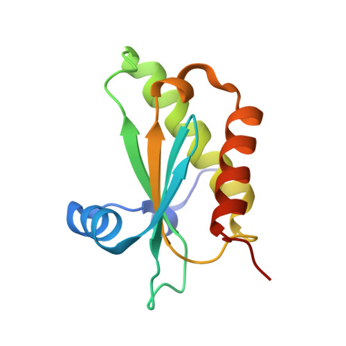
4JG4 - PubMed Abstract:
Coupled ligand binding and conformational change plays a central role in biological regulation. Ligands often regulate protein function by modulating conformational dynamics, yet the order in which binding and conformational change occurs are often hotly debated. Here we show that the "conformational selection versus induced fit" distinction on which this debate is based is a false dichotomy because the mechanism depends on ligand concentration. Using the binding of pyrophosphate (PPi) to Bacillus subtilis RNase P protein as a model, we show that coupled reactions are best understood as a change in flux between competing pathways with distinct orders of binding and conformational change. The degree of partitioning through each pathway depends strongly on PPi concentration, with ligand binding redistributing the conformational ensemble toward the folded state by both increasing folding rates and decreasing unfolding rates. These results indicate that ligand binding induces marked and varied changes in protein conformational dynamics, and that the order of binding and conformational change is ligand concentration dependent.
- Department of Biochemistry, Duke University Medical Center , Durham, North Carolina 27710, United States.
Organizational Affiliation: