4JFQ

A2 HLA complex with L8A heteroclitic variant of Melanoma peptide


Experimental Data Snapshot

  • Method: X-RAY DIFFRACTION
  • Resolution: 1.90 Å
  • R-Value Free: 0.297 
  • R-Value Work: 0.216 
  • R-Value Observed: 0.220 

wwPDB Validation   3D Report Full Report


This is version 1.3 of the entry. See complete history


Literature

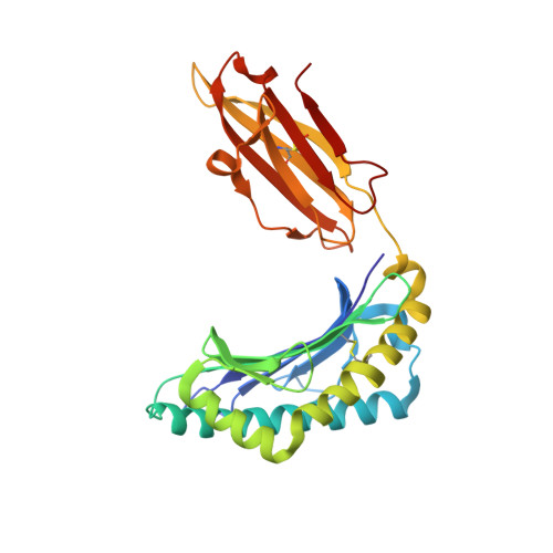
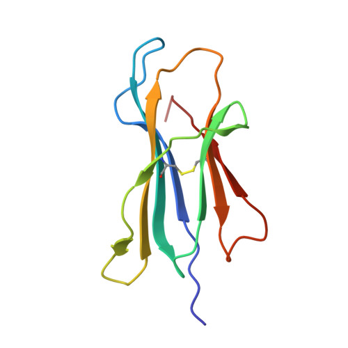
T-cell receptor specificity maintained by altered thermodynamics.

Madura, F.Rizkallah, P.J.Miles, K.M.Holland, C.J.Bulek, A.M.Fuller, A.Schauenburg, A.J.Miles, J.J.Liddy, N.Sami, M.Li, Y.Hossain, M.Baker, B.M.Jakobsen, B.K.Sewell, A.K.Cole, D.K.

(2013) J Biol Chem 288: 18766-18775

  • DOI: https://doi.org/10.1074/jbc.M113.464560
  • Primary Citation of Related Structures:  
    4JFD, 4JFE, 4JFF, 4JFH, 4JFO, 4JFP, 4JFQ

  • PubMed Abstract: 

    The T-cell receptor (TCR) recognizes peptides bound to major histocompatibility molecules (MHC) and allows T-cells to interrogate the cellular proteome for internal anomalies from the cell surface. The TCR contacts both MHC and peptide in an interaction characterized by weak affinity (KD = 100 nM to 270 μM). We used phage-display to produce a melanoma-specific TCR (α24β17) with a 30,000-fold enhanced binding affinity (KD = 0.6 nM) to aid our exploration of the molecular mechanisms utilized to maintain peptide specificity. Remarkably, although the enhanced affinity was mediated primarily through new TCR-MHC contacts, α24β17 remained acutely sensitive to modifications at every position along the peptide backbone, mimicking the specificity of the wild type TCR. Thermodynamic analyses revealed an important role for solvation in directing peptide specificity. These findings advance our understanding of the molecular mechanisms that can govern the exquisite peptide specificity characteristic of TCR recognition.


  • Organizational Affiliation

    Cardiff University School of Medicine, Heath Park, Cardiff CF14 4XN, United Kingdom.


Macromolecules
Find similar proteins by:  (by identity cutoff)  |  3D Structure
Entity ID: 1
MoleculeChains Sequence LengthOrganismDetailsImage
HLA class I histocompatibility antigen, A-2 alpha chain
A, D
276Homo sapiensMutation(s): 0 
Gene Names: HLA-AHLAA
UniProt & NIH Common Fund Data Resources
Find proteins for P04439 (Homo sapiens)
Explore P04439 
Go to UniProtKB:  P04439
PHAROS:  P04439
GTEx:  ENSG00000206503 
Entity Groups  
Sequence Clusters30% Identity50% Identity70% Identity90% Identity95% Identity100% Identity
UniProt GroupP04439
Sequence Annotations
Expand
  • Reference Sequence
Find similar proteins by:  (by identity cutoff)  |  3D Structure
Entity ID: 2
MoleculeChains Sequence LengthOrganismDetailsImage
Beta-2-microglobulin
B, E
100Homo sapiensMutation(s): 0 
Gene Names: B2MCDABP0092HDCMA22P
UniProt & NIH Common Fund Data Resources
Find proteins for P61769 (Homo sapiens)
Explore P61769 
Go to UniProtKB:  P61769
PHAROS:  P61769
GTEx:  ENSG00000166710 
Entity Groups  
Sequence Clusters30% Identity50% Identity70% Identity90% Identity95% Identity100% Identity
UniProt GroupP61769
Sequence Annotations
Expand
  • Reference Sequence

Find similar proteins by:  Sequence   |   3D Structure  

Entity ID: 3
MoleculeChains Sequence LengthOrganismDetailsImage
L8A heteroclitic Melanoma peptide
C, F
10N/AMutation(s): 0 
Entity Groups  
Sequence Clusters30% Identity50% Identity70% Identity90% Identity95% Identity100% Identity
Sequence Annotations
Expand
  • Reference Sequence
Small Molecules
Ligands 2 Unique
IDChains Name / Formula / InChI Key2D Diagram3D Interactions
MES
Query on MES

Download Ideal Coordinates CCD File 
N [auth A],
X [auth D]
2-(N-MORPHOLINO)-ETHANESULFONIC ACID
C6 H13 N O4 S
SXGZJKUKBWWHRA-UHFFFAOYSA-N
GOL
Query on GOL

Download Ideal Coordinates CCD File 
AA [auth E]
G [auth A]
H [auth A]
I [auth A]
J [auth A]
AA [auth E],
G [auth A],
H [auth A],
I [auth A],
J [auth A],
K [auth A],
L [auth A],
M [auth A],
O [auth B],
P [auth B],
Q [auth B],
R [auth B],
S [auth D],
T [auth D],
U [auth D],
V [auth D],
W [auth D],
Y [auth E],
Z [auth E]
GLYCEROL
C3 H8 O3
PEDCQBHIVMGVHV-UHFFFAOYSA-N
Experimental Data & Validation

Experimental Data

  • Method: X-RAY DIFFRACTION
  • Resolution: 1.90 Å
  • R-Value Free: 0.297 
  • R-Value Work: 0.216 
  • R-Value Observed: 0.220 
  • Space Group: P 1 21 1
Unit Cell:
Length ( Å )Angle ( ˚ )
a = 84.096α = 90
b = 58.359β = 109.82
c = 89.432γ = 90
Software Package:
Software NamePurpose
PHASERphasing
REFMACrefinement
PDB_EXTRACTdata extraction

Structure Validation

View Full Validation Report


Entry History 

Deposition Data

Revision History  (Full details and data files)

  • Version 1.0: 2013-05-29
    Type: Initial release
  • Version 1.1: 2013-06-05
    Changes: Database references
  • Version 1.2: 2013-06-19
    Changes: Database references
  • Version 1.3: 2013-07-17
    Changes: Database references