4JFG | pdb_00004jfg

Crystal structure of sfGFP-66-HqAla


Experimental Data Snapshot

  • Method: X-RAY DIFFRACTION
  • Resolution: 3.00 Å
  • R-Value Free: 
    0.312 (Depositor), 0.318 (DCC) 
  • R-Value Work: 
    0.259 (Depositor), 0.267 (DCC) 
  • R-Value Observed: 
    0.262 (Depositor) 

Starting Model: experimental
View more details

wwPDB Validation 3D Report Full Report

Validation slider image for 4JFG

This is version 2.1 of the entry. See complete history

Literature

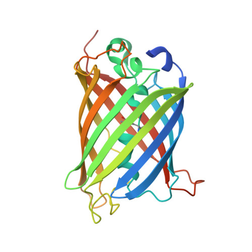
Significant expansion of the fluorescent protein chromophore through the genetic incorporation of a metal-chelating unnatural amino acid.

Liu, X.Li, J.Hu, C.Zhou, Q.Zhang, W.Hu, M.Zhou, J.Wang, J.

(2013) Angew Chem Int Ed Engl 52: 4805-4809

Macromolecule Content 

  • Total Structure Weight: 226.72 kDa 
  • Atom Count: 14,241 
  • Modeled Residue Count: 1,785 
  • Deposited Residue Count: 1,952 
  • Unique protein chains: 1

Macromolecules

Find similar proteins by:|  3D Structure
Entity ID: 1
MoleculeChains  Sequence LengthOrganismDetailsImage
Green fluorescent protein
A, B, C, D, E
A, B, C, D, E, F, G, H
244Aequorea victoriaMutation(s): 10 
Gene Names: GFP
UniProt
Find proteins for P42212 (Aequorea victoria)
Explore P42212 
Go to UniProtKB:  P42212
Entity Groups
Sequence Clusters30% Identity50% Identity70% Identity90% Identity95% Identity100% Identity
UniProt GroupP42212
Sequence Annotations
Expand
Reference Sequence

Small Molecules

Ligands 2 Unique
IDChains Name / Formula / InChI Key2D Diagram3D Interactions
HQY

Query on HQY


Download:Ideal Coordinates CCD File
DA [auth E]
GA [auth F]
I [auth A]
MA [auth G]
P [auth B]
DA [auth E],
GA [auth F],
I [auth A],
MA [auth G],
P [auth B],
SA [auth H],
U [auth C],
Z [auth D]
quinolin-8-ol
C9 H7 N O
MCJGNVYPOGVAJF-UHFFFAOYSA-N
CS

Query on CS


Download:Ideal Coordinates CCD File
AA [auth D]
BA [auth D]
CA [auth D]
EA [auth E]
FA [auth E]
AA [auth D],
BA [auth D],
CA [auth D],
EA [auth E],
FA [auth E],
HA [auth F],
IA [auth F],
J [auth A],
JA [auth F],
K [auth A],
KA [auth F],
L [auth A],
LA [auth F],
M [auth A],
N [auth A],
NA [auth G],
O [auth A],
OA [auth G],
PA [auth G],
Q [auth B],
QA [auth G],
R [auth B],
RA [auth G],
S [auth B],
T [auth B],
TA [auth H],
UA [auth H],
V [auth C],
W [auth C],
X [auth C],
Y [auth C]
CESIUM ION
Cs
NCMHKCKGHRPLCM-UHFFFAOYSA-N
Modified Residues  1 Unique
IDChains TypeFormula2D DiagramParent
KWS
Query on KWS
A, B, C, D, E
A, B, C, D, E, F, G, H
L-PEPTIDE LINKINGC9 H13 N3 O4THR, SER, GLY

Experimental Data & Validation

Experimental Data

  • Method: X-RAY DIFFRACTION
  • Resolution: 3.00 Å
  • R-Value Free:  0.312 (Depositor), 0.318 (DCC) 
  • R-Value Work:  0.259 (Depositor), 0.267 (DCC) 
  • R-Value Observed: 0.262 (Depositor) 
Space Group: P 21 21 21
Unit Cell:
Length ( Å )Angle ( ˚ )
a = 88.174α = 90
b = 148.901β = 90
c = 161.35γ = 90
Software Package:
Software NamePurpose
HKL-2000data collection
MLPHAREphasing
PHENIXrefinement
HKL-2000data reduction
HKL-2000data scaling

Structure Validation

View Full Validation Report


Entry History 

Deposition Data

Revision History  (Full details and data files)

  • Version 1.0: 2013-10-02
    Type: Initial release
  • Version 1.1: 2023-11-08
    Changes: Data collection, Database references, Derived calculations, Refinement description
  • Version 2.0: 2023-11-15
    Changes: Atomic model, Data collection, Derived calculations
  • Version 2.1: 2026-03-18
    Changes: Structure summary