4JF7 | pdb_00004jf7

Structure of the parainfluenza virus 5 (PIV5) hemagglutinin-neuraminidase (HN) ectodomain


Experimental Data Snapshot

  • Method: X-RAY DIFFRACTION
  • Resolution: 2.50 Å
  • R-Value Free: 
    0.207 (Depositor), 0.204 (DCC) 
  • R-Value Work: 
    0.166 (Depositor), 0.165 (DCC) 
  • R-Value Observed: 
    0.166 (Depositor) 

wwPDB Validation 3D Report Full Report

Validation slider image for 4JF7

Ligand Structure Quality Assessment 


This is version 2.1 of the entry. See complete history

Literature

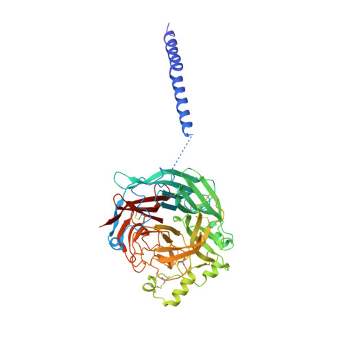
Structure of the Parainfluenza Virus 5 (PIV5) Hemagglutinin-Neuraminidase (HN) Ectodomain.

Welch, B.D.Yuan, P.Bose, S.Kors, C.A.Lamb, R.A.Jardetzky, T.S.

(2013) PLoS Pathog 9: e1003534-e1003534

  • DOI: https://doi.org/10.1371/journal.ppat.1003534
  • Primary Citation Related Structures: 
    4JF7

  • PubMed Abstract: 

    Paramyxoviruses cause a wide variety of human and animal diseases. They infect host cells using the coordinated action of two surface glycoproteins, the receptor binding protein (HN, H, or G) and the fusion protein (F). HN binds sialic acid on host cells (hemagglutinin activity) and hydrolyzes these receptors during viral egress (neuraminidase activity, NA). Additionally, receptor binding is thought to induce a conformational change in HN that subsequently triggers major refolding in homotypic F, resulting in fusion of virus and target cell membranes. HN is an oligomeric type II transmembrane protein with a short cytoplasmic domain and a large ectodomain comprising a long helical stalk and large globular head domain containing the enzymatic functions (NA domain). Extensive biochemical characterization has revealed that HN-stalk residues determine F specificity and activation. However, the F/HN interaction and the mechanisms whereby receptor binding regulates F activation are poorly defined. Recently, a structure of Newcastle disease virus (NDV) HN ectodomain revealed the heads (NA domains) in a "4-heads-down" conformation whereby two of the heads form a symmetrical interaction with two sides of the stalk. The interface includes stalk residues implicated in triggering F, and the heads sterically shield these residues from interaction with F (at least on two sides). Here we report the x-ray crystal structure of parainfluenza virus 5 (PIV5) HN ectodomain in a "2-heads-up/2-heads-down" conformation where two heads (covalent dimers) are in the "down position," forming a similar interface as observed in the NDV HN ectodomain structure, and two heads are in an "up position." The structure supports a model in which the heads of HN transition from down to up upon receptor binding thereby releasing steric constraints and facilitating the interaction between critical HN-stalk residues and F.


  • Organizational Affiliation
    • Department of Molecular Biosciences, Northwestern University, Evanston, Illinois, USA.

Macromolecule Content 

  • Total Structure Weight: 230.93 kDa 
  • Atom Count: 16,400 
  • Modeled Residue Count: 1,987 
  • Deposited Residue Count: 2,052 
  • Unique protein chains: 1

Macromolecules

Find similar proteins by:|  3D Structure
Entity ID: 1
MoleculeChains  Sequence LengthOrganismDetailsImage
Hemagglutinin-neuraminidaseA [auth D],
B [auth A],
C [auth B],
D [auth C]
513Simian virus 5 (strain W3)Mutation(s): 0 
Gene Names: HN
EC: 3.2.1.18
UniProt
Find proteins for P04850 (Parainfluenza virus 5 (strain W3))
Explore P04850 
Go to UniProtKB:  P04850
Entity Groups
Sequence Clusters30% Identity50% Identity70% Identity90% Identity95% Identity100% Identity
UniProt GroupP04850
Glycosylation
Glycosylation Sites: 3
Sequence Annotations
Expand
Reference Sequence

Oligosaccharides

Help  
Entity ID: 2
MoleculeChains Length2D Diagram GlycosylationD Interactions
alpha-D-mannopyranose-(1-4)-2-acetamido-2-deoxy-beta-D-glucopyranose-(1-4)-2-acetamido-2-deoxy-beta-D-glucopyranose
E, F
3N-Glycosylation
Glycosylation Resources
GlyTouCan: G62182OO
GlyCosmos: G62182OO
GlyGen: G62182OO
Entity ID: 3
MoleculeChains Length2D Diagram GlycosylationD Interactions
2-acetamido-2-deoxy-beta-D-glucopyranose-(1-4)-2-acetamido-2-deoxy-beta-D-glucopyranose
G, H, I, J
2N-Glycosylation
Glycosylation Resources
GlyTouCan: G42666HT
GlyCosmos: G42666HT
GlyGen: G42666HT

Small Molecules

Ligands 3 Unique
IDChains Name / Formula / InChI Key2D Diagram3D Interactions
NAG

Query on NAG


Download:Ideal Coordinates CCD File
M [auth D]
O [auth A]
Q [auth A]
T [auth B]
W [auth C]
M [auth D],
O [auth A],
Q [auth A],
T [auth B],
W [auth C],
X [auth C]
2-acetamido-2-deoxy-beta-D-glucopyranose
C8 H15 N O6
OVRNDRQMDRJTHS-FMDGEEDCSA-N
SO4

Query on SO4


Download:Ideal Coordinates CCD File
L [auth D],
P [auth A],
S [auth B],
V [auth C]
SULFATE ION
O4 S
QAOWNCQODCNURD-UHFFFAOYSA-L
CA

Query on CA


Download:Ideal Coordinates CCD File
K [auth D],
N [auth A],
R [auth B],
U [auth C]
CALCIUM ION
Ca
BHPQYMZQTOCNFJ-UHFFFAOYSA-N

Experimental Data & Validation

Experimental Data

  • Method: X-RAY DIFFRACTION
  • Resolution: 2.50 Å
  • R-Value Free:  0.207 (Depositor), 0.204 (DCC) 
  • R-Value Work:  0.166 (Depositor), 0.165 (DCC) 
  • R-Value Observed: 0.166 (Depositor) 
Space Group: I 4
Unit Cell:
Length ( Å )Angle ( ˚ )
a = 194.39α = 90
b = 194.39β = 90
c = 185.979γ = 90
Software Package:
Software NamePurpose
HKL-2000data collection
PHENIXrefinement
HKL-2000data reduction
HKL-2000data scaling

Structure Validation

View Full Validation Report


Ligand Structure Quality Assessment 


Entry History 

Deposition Data

Revision History  (Full details and data files)

  • Version 1.0: 2013-09-04
    Type: Initial release
  • Version 2.0: 2020-07-29
    Type: Remediation
    Reason: Carbohydrate remediation
    Changes: Atomic model, Data collection, Database references, Derived calculations, Structure summary
  • Version 2.1: 2024-11-06
    Changes: Data collection, Database references, Structure summary