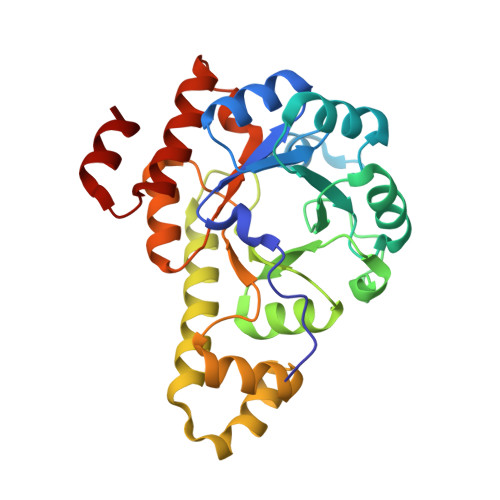
4JDY

Crystal structure of Rv2606c


Experimental Data Snapshot

  • Method: X-RAY DIFFRACTION
  • Resolution: 1.80 Å
  • R-Value Free: 0.211 
  • R-Value Work: 0.183 
  • R-Value Observed: 0.184 

Starting Model: experimental
View more details

wwPDB Validation   3D Report Full Report


This is version 1.3 of the entry. See complete history


Literature

Crystal structure of Mycobacterium tuberculosis Rv2606c: a pyridoxal biosynthesis lyase.

Kim, S.Kim, K.J.

(2013) Biochem Biophys Res Commun 435: 255-259

  • DOI: https://doi.org/10.1016/j.bbrc.2013.04.068
  • Primary Citation of Related Structures:  
    4JDY

  • PubMed Abstract: 

    Tuberculosis is a lethal infectious disease caused by Mycobacterium tuberculosis. We determined the crystal structure of Rv2606c, a potential pyridoxal biosynthesis lyase (PdxS), from M. tuberculosis H37Rv at 1.8 Å resolution. The overall structure of the protein, composed of a (β/α)8-barrel and two small 310-helices, was quite similar to those of other PdxS proteins. A glycerol molecule was observed to be bound at the active site of the Rv2606c structure through interactions with the conserved residues of Asp29 and Lys86, providing information regarding the potential active site and the substrate-binding environment of the protein. The interface for Rv2606c dodecamerization, which is primarily mediated by salt bridges and hydrophobic interactions, was quite different from those of other PdxS proteins. Furthermore, we observed that the Rv2606c and Rv2604c form a stable complex, suggesting that these proteins might function as PdxS and PdxT in M. tuberculosis.


  • Organizational Affiliation

    School of Life Science and Biotechnology, Kyungpook National University, Republic of Korea.


Macromolecules
Find similar proteins by:  (by identity cutoff)  |  3D Structure
Entity ID: 1
MoleculeChains Sequence LengthOrganismDetailsImage
Pyridoxal biosynthesis lyase PdxS
A, B, C
264Mycobacterium tuberculosisMutation(s): 0 
Gene Names: pdxSRv2606cMT2681MTCY1A10.27
UniProt
Find proteins for P9WII9 (Mycobacterium tuberculosis (strain ATCC 25618 / H37Rv))
Explore P9WII9 
Go to UniProtKB:  P9WII9
Entity Groups  
Sequence Clusters30% Identity50% Identity70% Identity90% Identity95% Identity100% Identity
UniProt GroupP9WII9
Sequence Annotations
Expand
  • Reference Sequence
Experimental Data & Validation

Experimental Data

  • Method: X-RAY DIFFRACTION
  • Resolution: 1.80 Å
  • R-Value Free: 0.211 
  • R-Value Work: 0.183 
  • R-Value Observed: 0.184 
  • Space Group: I 2 2 2
Unit Cell:
Length ( Å )Angle ( ˚ )
a = 110.752α = 90
b = 126.08β = 90
c = 180.816γ = 90
Software Package:
Software NamePurpose
DENZOdata reduction
SCALEPACKdata scaling
MOLREPphasing
REFMACrefinement
PDB_EXTRACTdata extraction
HKL-2000data collection
HKL-2000data reduction
HKL-2000data scaling

Structure Validation

View Full Validation Report


Entry History 

Deposition Data

Revision History  (Full details and data files)

  • Version 1.0: 2013-05-22
    Type: Initial release
  • Version 1.1: 2017-11-15
    Changes: Refinement description
  • Version 1.2: 2022-08-24
    Changes: Database references, Derived calculations
  • Version 1.3: 2023-11-08
    Changes: Data collection, Refinement description